Immuno-oncology Assays
Antibody-dependent cellular phagocytosis (ADCP)
Antibody-dependent cellular phagocytosis is a mechanism by which antibody-opsonized target cells activate the FcγRs on the surface of monocytes/macrophages or engineered immune cells to induce phagocytosis, resulting in the internalization and degradation of the target cell through acidification of the phagosome.
- Examine ADCP activity of your proprietary therapeutic antibody using physiologically relevant in vitro model
- Bespoke ADCP assays using primary monocytes/macrophages from multiple donors or genetically engineered immune cell lines
- Develop cell lines expressing target antigen of your interest
Antibody-Dependent Cellular Phagocytosis (ADCP)
THP-1 monocytic cell line derived macrophages - ADCP
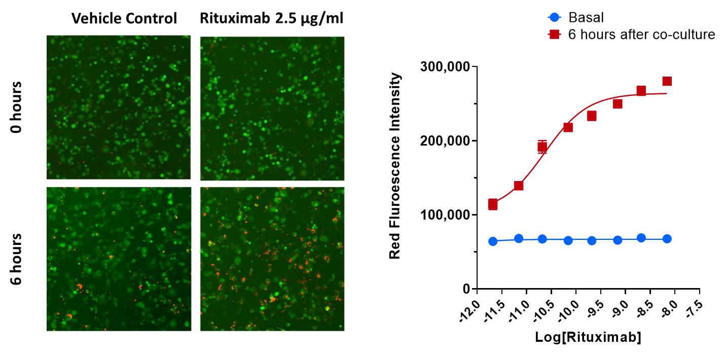
Target Raji cells were pre-treated with or without Rituximab at different concentrations and stained with pHrodo Red (red). THP-1 derived macrophages were stained with calcein (green). To assess antibody-dependent cellular phagocytosis of target Raji cells by THP-1 derived macrophages, the two cell types were co-cultured for 6 hours. Data is represented as the red fluorescence intensity (RFI) at basal (0 hours) vs. 6 hours in all treatment groups.
Primary monocyte derived macrophages - ADCP
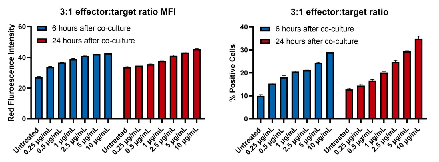
The phagocytic activity of primary monocyte-derived macrophages towards MCF-7 breast cancer cells treated ± HER2 targeting monoclonal antibody Trastuzumab. MCF-7 breast cancer cells were treated with different concentrations of Trastuzumab for 1 hour prior to initiating the co-culture with primary-derived macrophages at an effector to target ratio of 3:1. Staining of MCF-7 cells with Incucyte® phRodo® dye produces red fluorescent signals as a result of phagocytosis in response to the treatment conditions (A). The percentage of target cells positive for phagocytosis was determined by dividing the number of target cells (MCF-7) that have been phagocytosed relative to the total number of target cells (MCF-7) seeded at the start of co-culture (B). These two parameters were assessed at 0 and 6 hours post co-culture. Data was represented as the mean ± SEM of 3 replicates.
Request a consultation with Cellomatics Biosciences today
Our experienced team of in vitro laboratory scientists will work with you to understand your project and provide a bespoke project plan with a professional, flexible service and a fast turnaround time.
To request a consultation where we can discuss your exact requirements, please contact Cellomatics Biosciences.


