

The shift to utilising 3D cancer models remains an attractive option to investigate more clinically relevant features. We have extensive experience producing spheroids from a wide range of cell type; breast, colon, lung, liver and brain. Following drug treatment, fluorescently labelled dyes enabled a detailed annotation of the spheroids to delineate regions that are either viable or have undergone cell death. Alternatively, we can quantify spheroid area with brightfield images or assess cytotoxicity using the CellTiter Glo 3D assay. In addition we have developed models to track immune cell invasion into spheroids
Complementing this work is our two imaging platforms, JuLI Stage and ImageXpress Pico System, which can perform an array of functions including live imaging and time lapse capture to support with spheroid assays and related projects.
To find out how Cellomatics can support your spheroid assays and project requirements, request a quote below.
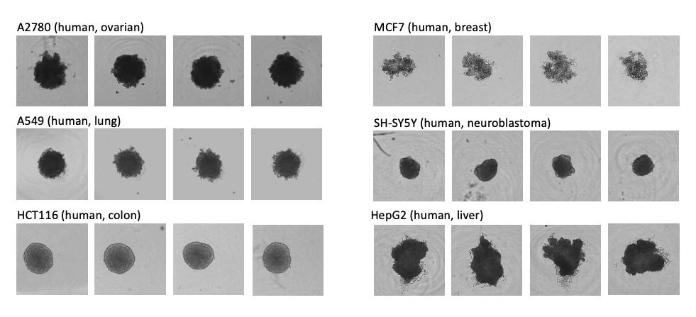
Spheroid formation across a panel of cell lines representing different cancer models. Spheroids were grown in ULA (ultra-low adhesion)-U-bottomed, 96-well culture plates. At 72 h post-seeding, spheroids were imaged using a JuliStage platform fitted with a x4 objective confirming that each well contained a single spheroid; comparison of multiple spheroids confirms that morphology and size were consistent.
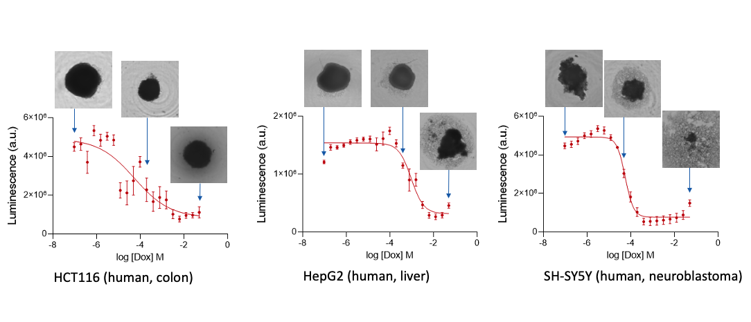
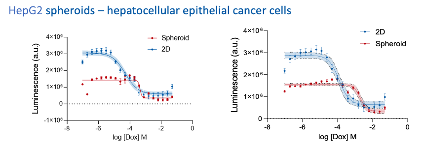
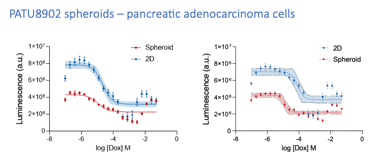
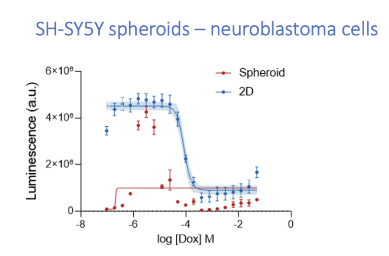
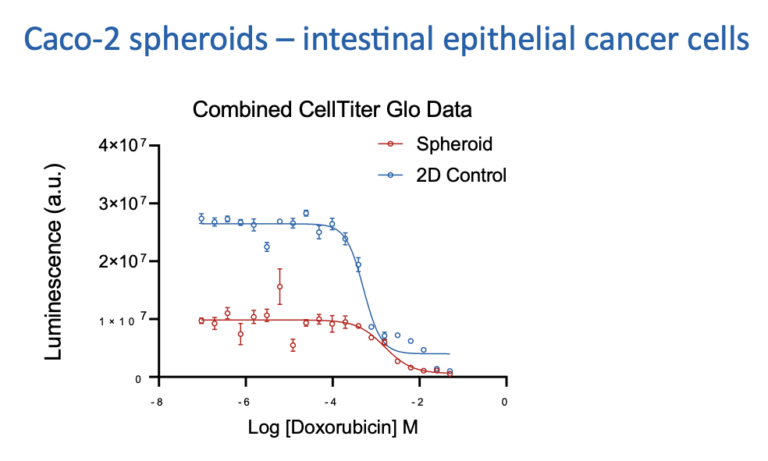
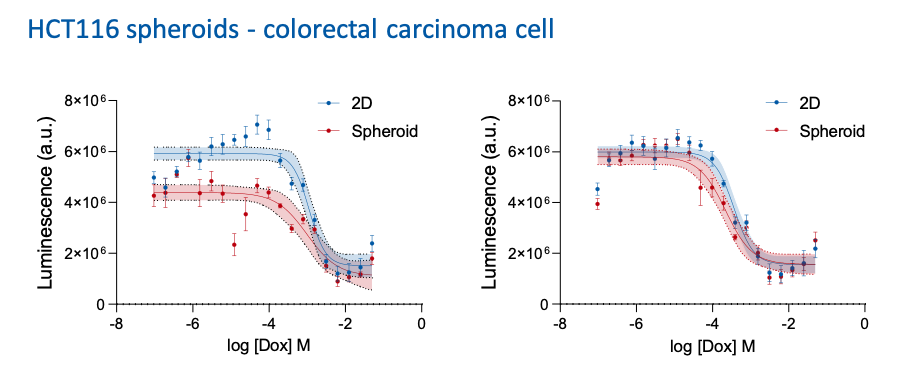
Effects of doxorubicin on spheroid viability. Spheroids were subsequently incubated for 9 days in the presence of the chemotherapeutic agent Doxorubicin (media + Dox was replaced at day 4 post-initiation of treatment). Cell viability was assessed by means of the Promega CellTiter Glo-3D assay kit, and dose-response curves allowing the determination of pEC50 values were modelled in GraphPad Prism (n=8 per condition ±S.E.M.). N=8 across 2 runs of 4.
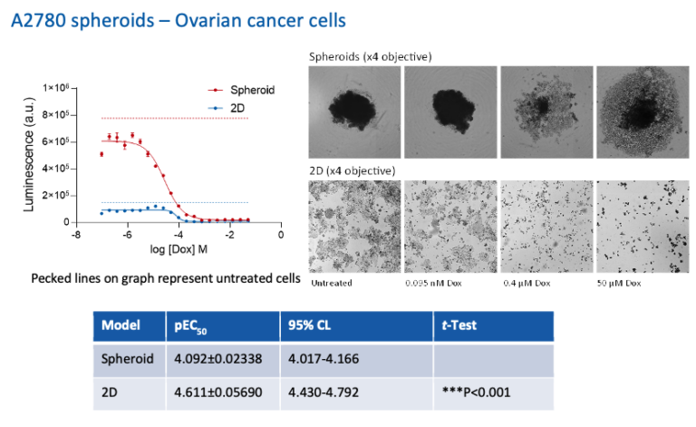
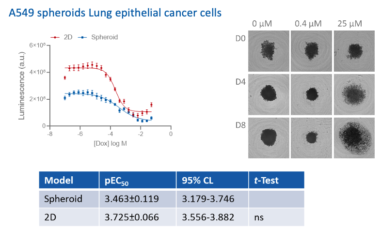
A concentration response to Doxorubin was assessed by culturing cancer cells in 2D or 3D speheroid models. Spheroids were formed for at least 72 h prior to treatment followed by doxorubicin treatment for 7 days. Cell Tite Glo assay was performed to determing cell viability and is expressed as relative luminescence.
Our experienced team of in vitro laboratory scientists will work with you to understand your project and provide a bespoke project plan with a professional, flexible service and a fast turnaround time.
To request a consultation where we can discuss your exact requirements, please contact Cellomatics Biosciences.
Cellomatics Biosciences Limited
10 Colwick Quays Business Park
Road No2, Colwick Nottingham NG4 2JY, UK
+44 (115) 865 4101
info@cellomaticsbio.com

Cellomatics Biosciences Limited
10 Colwick Quays Business Park
Road No2, Colwick Nottingham NG4 2JY, UK
+44 (115) 865 4101
info@cellomaticsbio.com


| Cookie | Duration | Description |
|---|---|---|
| cookielawinfo-checkbox-analytics | 11 months | This cookie is set by GDPR Cookie Consent plugin. The cookie is used to store the user consent for the cookies in the category "Analytics". |
| cookielawinfo-checkbox-functional | 11 months | The cookie is set by GDPR cookie consent to record the user consent for the cookies in the category "Functional". |
| cookielawinfo-checkbox-necessary | 11 months | This cookie is set by GDPR Cookie Consent plugin. The cookies is used to store the user consent for the cookies in the category "Necessary". |
| cookielawinfo-checkbox-others | 11 months | This cookie is set by GDPR Cookie Consent plugin. The cookie is used to store the user consent for the cookies in the category "Other. |
| cookielawinfo-checkbox-performance | 11 months | This cookie is set by GDPR Cookie Consent plugin. The cookie is used to store the user consent for the cookies in the category "Performance". |
| viewed_cookie_policy | 11 months | The cookie is set by the GDPR Cookie Consent plugin and is used to store whether or not user has consented to the use of cookies. It does not store any personal data. |