Inflammatory Bowel Disease (IBD)
IBD is a term used to describe disorders that involve chronic/recurrent inflammation of the colon and small intestine.
Ulcerative colitis is mainly localised into the colon (large intestine), while Crohn’s disease can affect any part of the digestive system. In both cases, symptoms may range from mild to severe, with periods of active illness followed by temporary remission. There is currently no cure for IBD, and medications tend to reduce the symptoms preventing severe complications, which include colon cancer.
Causes:
- Defects in the intestinal barrier
- Impaired immune function
- Genetic predispositions (e.g. NOD2 mutation)
- Environmental factors
Characteristics:
- Increased proinflammatory immune response
- Increased permeability of the intestinal epithelial barrier
- Increased mucus production
- Rearrangement of tight junction proteins
In vitro models –
Relevant cell types:
- Caco-2 cells – Human Colorectal adenocarcinoma epithelial-like cell line
- InEpC – Primary Human Intestinal Epithelial Cells
- Immune cells – Freshly isolated from peripheral blood of healthy/diseased volunteers


Caco2 Transwell mono-culture IBD model
Caco-2 Model for IBD: Tight Junction Proteins
Caco-2 cells were differentiated for 21 days and stained for tight junction proteins such as ZO-1 (purple) and Occludin (orange) along with DAPI (blue– nucleus). The images were captured at 20X magnification using high content imager and the intensities of each fluorescence analysed.
Caco-2 Model for IBD: TEER Reported
Caco-2 cells were seeded onto trans-well inserts and allowed to differentiate for 21 days and TEER was measured regularly. On day 21 (Basal), the monolayer was treated Dexamethasone and stimulated with LPS+ IL1beta cocktail for 48 hours. TEER measurements were continued at 24 and 48 hours post treatment (pt) and reported as Ω.cm2 (n=3±SEM).
Caco-2 Model for IBD: Tight Junction Proteins
Caco-2 cells differentiated for 21 days were treated with Dexamethasone followed by stimulation with LPS+ IL1beta cocktail for 48 hours. The monolayers were stained for tight junction protein ZO-1 (green) with DAPI (blue – nucleus). The images were captured using ImageXpress Pico and the fluorescent intensities analysed to obtain Percentage (%) of Positive Cells.
Caco-2 Model for IBD: Permeability
Caco-2 cells were differentiated for 21 days and treated with Dexamethasone followed by stimulation with LPS+ IL1beta cocktail for 48 hours. FITC-Dextran was added to the apical chamber along with the stimulants. Breaks in the monolayer were detected by the permeability of FITC-Dextran into the basal chamber. The RFU of all the conditions were normalised to the untreated control (100). A significant reduction in FITC dextran was observed in Dexamethasone when compared to stimulated condition (****p<0.0001; n=3±SEM).
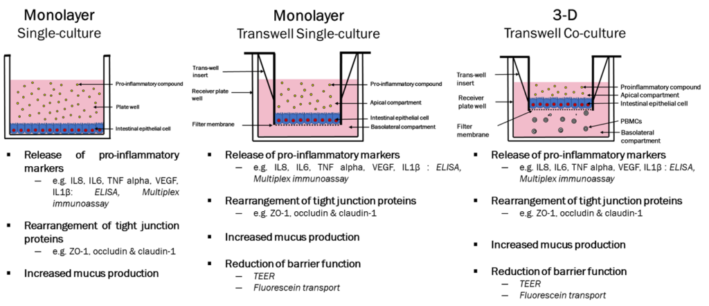
Caco-2 Model for IBD: Inflammatory Mediators
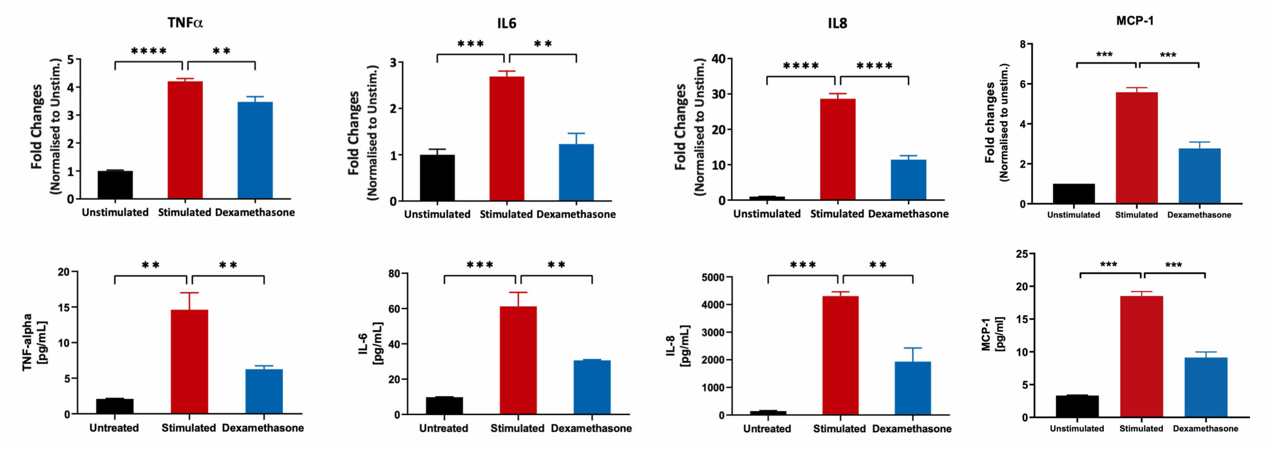
Caco-2 cells were differentiated for 21 days and subsequently treated with Dexamethasone, followed by stimulation with LPS+ IL1beta cocktail. mRNA expression in cell lysates was quantified using the Luminex Quantigene™ Assay. The supernatants were analysed for inflammatory mediators using a Luminex Multiplex Assay (**p<0.01; ***p<0.001; ****p<0.0001; n=3 mean±SEM).
Caco2/THP-1 3D transwell co-culture model
Caco2/THP-1 co-culture IBD model


Caco-2 cells differentiated for 21 days and PMA differentiated THP-1 cells were grown in co-culture and stimulated with LPS and IL-1β for 48hrs in the presence or absence of Sulfasalazine. The monolayers were stained after 48hrs for tight junction protein ZO-1 (pink) with DAPI (blue– nucleus). The images show a reduction in the ZO-1 protein in the stimulated cells compared to the well defined protein structure seen in the unstimulated cells. Also, pre-treatment with sulfasalazine prevented damage to the tight junction protein when compared to the stimulated cells. Images were captured using ImageXpress Pico at a x20 magnification.
Caco-2/THP-1 co-culture 3D Model for IBD: TEER Reported


Caco-2 cells differentiated for 21 days and PMA differentiated THP-1 cells were grown in co-culture and stimulated with LPS and IL-1β for 48hrs to induce an Inflammatory Bowel Disease phenotype in the presence or absence of Sulfasalazine and 5-Aminosalicylic Acid. The TEER reported for all the conditions (compounds and time of treatment) were normalised to the TEER reported on day 21 of differentiation (Basal) for each of the transwells. This data was analysed for both compounds using Two-way ANOVA followed by Dunnett’s multiple comparisons test [95% CI of diff.] (*p<0.05, **p<0.01,****p<0.0001). For each time point, all conditions were compared to the Stimulated control. The co-culture model treated with Sulfasalazine, was significantly different at 48hours and showed an improvement in TEER values when compared to the stimulated co-culture model.
Note: Unstimulated = Co-culture (Caco-2 +THP-1) alone, Stimulated= Co-culture (Caco-2 +THP-1) + LPS + IL-1β.
Pro-inflammatory marker suppression by Sulfasalazine and 5-Aminosalicylic Acid


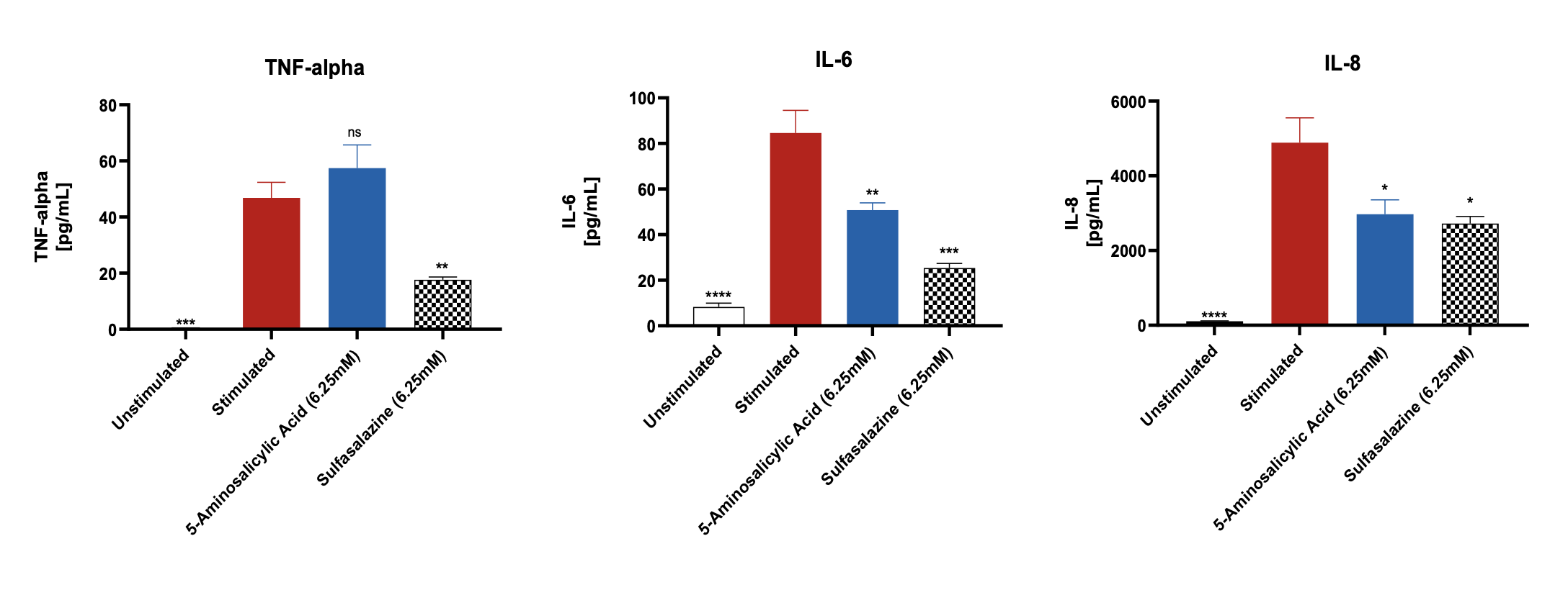
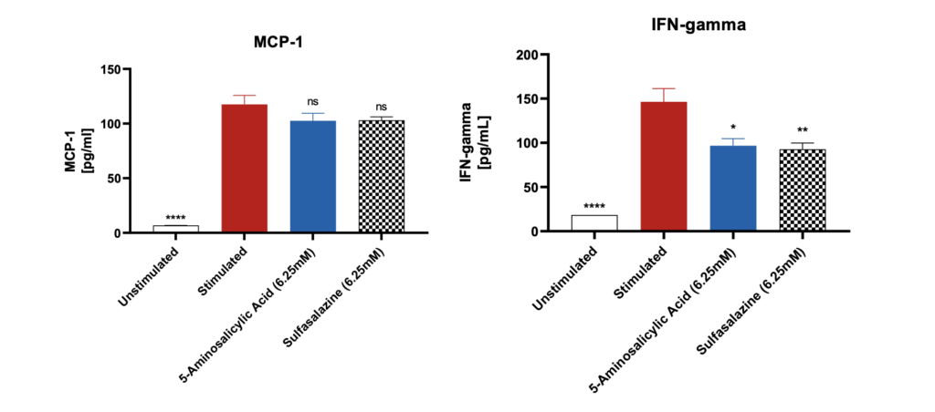
Significantly increased levels of pro-inflammatory markers were observed after stimulation of the differentiated Caco-2 monolayers with IL-1β and LPS when compared to unstimulated control. The levels of these markers were suppressed when the Caco-2 monolayers were pre-treated with the reference compounds. Data was analyzed using Ordinary One-way ANOVA followed by Dunnett’s multiple comparisons test [95% CI of diff.] (*p<0.05, **p<0.01, ***p<0.001, ****p<0.0001, ns= not significant). All the conditions were compared to the stimulated control for each of the markers and showed significant differences.
Note: Unstimulated = Co-culture (Caco-2 +THP-1), Stimulated= Co-culture (Caco-2 +THP-1) + LPS + IL-1β.
Lactate Dehydrogenase (LDH) release by Caco-2 cells


Data was analyzed using Ordinary One-way ANOVA followed by Dunnett’s multiple comparisons test [95% CI of diff.] (*p<0.05, **p<0.01, ***p<0.001, ****p<0.0001, ns= not significant). The LDH positive control, provided with the assay, generated significant OD 450 values (p<0.0001) compared to the unstimulated control. Both Sulfasalazine and 5-Aminosalicylic Acid showed no significant LDH release when compared to the unstimulated control.
Request a consultation with Cellomatics Biosciences today
Our experienced team of in vitro laboratory scientists will work with you to understand your project and provide a bespoke project plan with a professional, flexible service and a fast turnaround time.
To request a consultation where we can discuss your exact requirements, please contact Cellomatics Biosciences.