
An inflammatory skin disorder, triggered or exacerbated by a number of genetic, environmental, or immunological factors. Characterised by
We offer options of different cell-based approaches for our clients to choose from:
Primary human Keratinocyte/Immortalised keratinocyte cell line & blood cell models – monoculture or co-culture
o Readout: IL8, GRO-alpha, IL6, IL4, IL22, IL12, IL23, TSLP, GM-CSF, IFN gamma
o Read out: BrdU, MTT, Alamar Blue, Ki-67, Caspase3/7
o Read out: mRNA levels of epidermal differentiation and tight junction protein markers
Keratinocyte differentiation was assessed following treatment with calcium + serum, treatment with a cytokine cocktail, or as a consequence of reaching high confluence (untreated). The majority of untreated control cells differentiated by day 6. Differentiated cells could be readily identified by their large size and cobblestone morphology (white arrows). Treatment with a combination of calcium and serum caused the keratinocytes to rapidly differentiate (>24 hours), resulting in flattened, fibrous, skin-like morphology which was sustained throughout the 6 days of treatment. The cytokine-treated cells (exposed to TNF-α, Oncostatin-M and IL1-α) seemed to exbibit delayed, aberrant differentiation, which is one of the hallmarks of psoriasis. Black arrows highlight irregular filament formation visible only in cytokine treated cells.
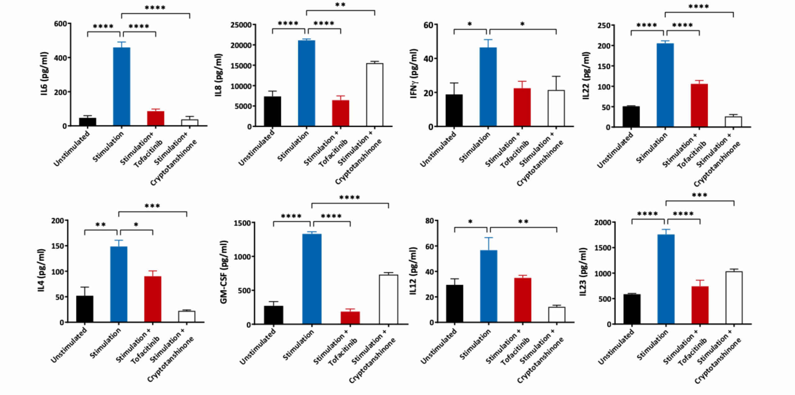
Human epidermal keratinocytes were differentiated in media supplemented with FBS and Calcium. The cells were then pre-treated with JAK3/STAT3 inhibitors followed by stimulation with a cytokine cocktail of IL1α + TNFα + Oncostatin M for 48 hours. Supernatants were analysed for various inflammatory mediators using Luminex Multiplex Assay (n=3±SEM;*p<0.05; **p<0.01; ***p<0.001; ****p<0.0001)
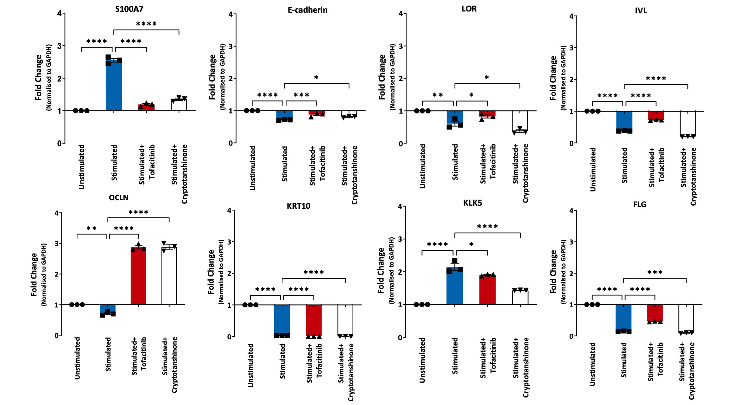
Human epidermal keratinocytes were allowed to differentiate in media supplemented with FBS and Calcium. The cells were then pre-treated with JAK3/STAT3 inhibitors followed by stimulation with a cytokine cocktail of IL1α + TNFα + Oncostatin M for 48 hours. Gene expression levels of key differentiation markers were analysed using Luminex Quantigene™ Assay. The mRNA expressions are normalised to GAPDH and fold change calculated relative to the unstimulated control (n=3±SEM; *p<0.05;**p<0.01;***p<0.001; ****p<0.0001).
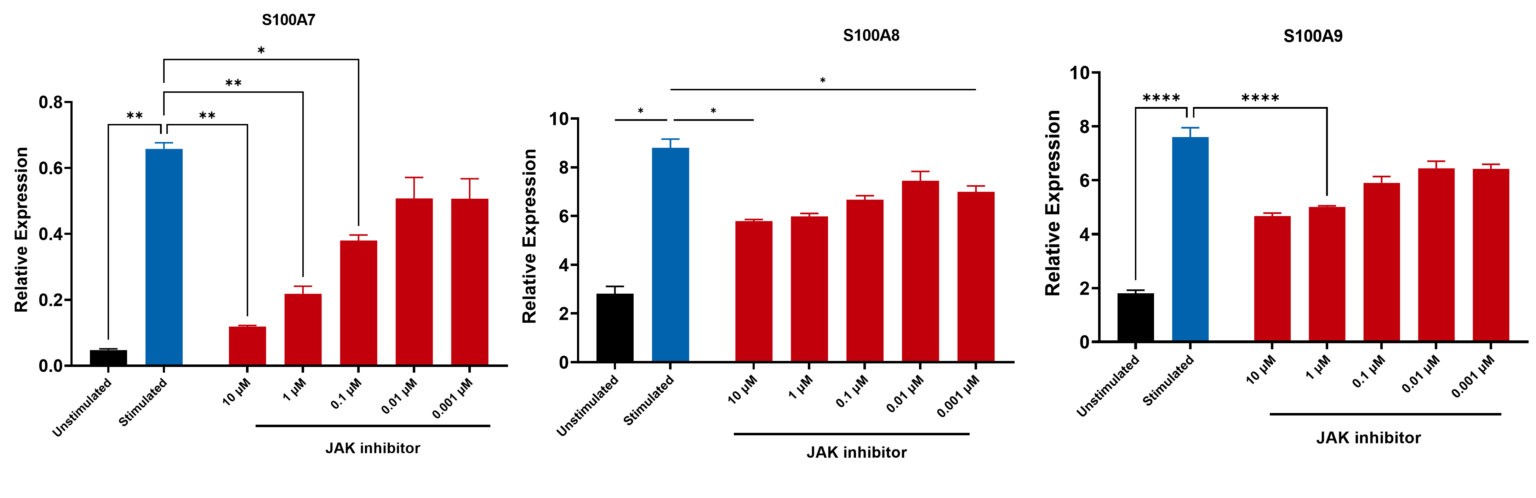
Human epidermal keratinocytes were allowed to differentiate. The cells were then pre-treated with JAK3 inhibitors followed by stimulation with a cytokine cocktail of IL1α + TNFα + Oncostatin M for 48 hours. Gene expression levels of key differentiation markers were analysed using Luminex Quantigene™ Assay. The mRNA expressions are normalised to GAPDH and fold change calculated relative to the unstimulated control (n=3±SEM; *p<0.05;**p<0.01;***p<0.001; ****p<0.0001).
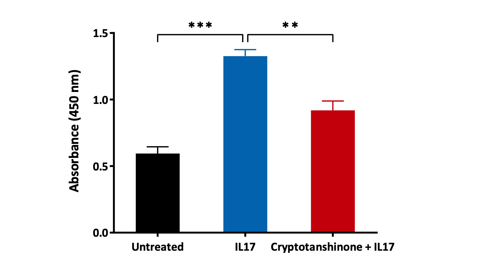
Healthy Human epidermal keratinocytes were stimulated with recombinant IL17A and cultured in the presence or absence of STAT3 Inhibitor (Cryptotanshinone) for 5 days. A BrdU incorporation assay was then performed and results are expressed as absorbance values (n=3, mean±SEM; **p<0.01; ***p<0.001).
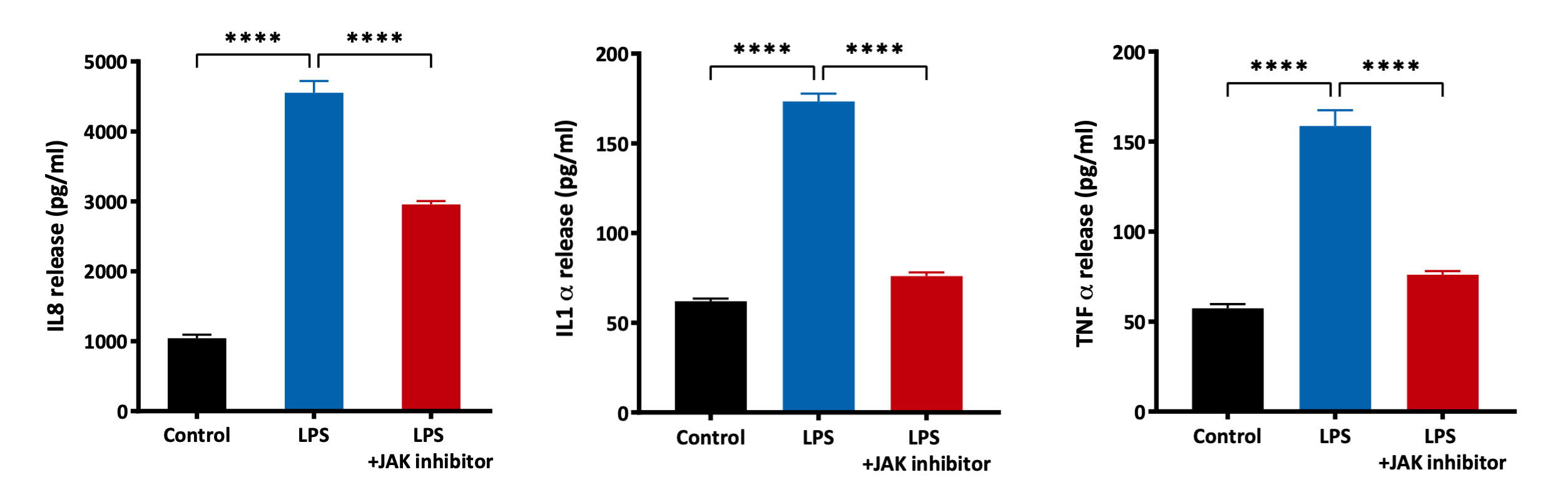
Effect of JAK Inhibitor on LPS-induced IL8, TNFα and IL1α release from primary human epidermal keratinocytes after 48 hours of treatment. The analytes were measured in cell supernatants using a Luminex Multiplex Assay (n=3, mean±SEM; ****p<0.0001).
Our experienced team of in vitro laboratory scientists will work with you to understand your project and provide a bespoke project plan with a professional, flexible service and a fast turnaround time.
To request a consultation where we can discuss your exact requirements, please contact Cellomatics Biosciences.
Cellomatics Biosciences Limited
10 Colwick Quays Business Park
Road No2, Colwick Nottingham NG4 2JY, UK
+44 (115) 865 4101
info@cellomaticsbio.com

Cellomatics Biosciences Limited
10 Colwick Quays Business Park
Road No2, Colwick Nottingham NG4 2JY, UK
+44 (115) 865 4101
info@cellomaticsbio.com


| Cookie | Duration | Description |
|---|---|---|
| cookielawinfo-checkbox-analytics | 11 months | This cookie is set by GDPR Cookie Consent plugin. The cookie is used to store the user consent for the cookies in the category "Analytics". |
| cookielawinfo-checkbox-functional | 11 months | The cookie is set by GDPR cookie consent to record the user consent for the cookies in the category "Functional". |
| cookielawinfo-checkbox-necessary | 11 months | This cookie is set by GDPR Cookie Consent plugin. The cookies is used to store the user consent for the cookies in the category "Necessary". |
| cookielawinfo-checkbox-others | 11 months | This cookie is set by GDPR Cookie Consent plugin. The cookie is used to store the user consent for the cookies in the category "Other. |
| cookielawinfo-checkbox-performance | 11 months | This cookie is set by GDPR Cookie Consent plugin. The cookie is used to store the user consent for the cookies in the category "Performance". |
| viewed_cookie_policy | 11 months | The cookie is set by the GDPR Cookie Consent plugin and is used to store whether or not user has consented to the use of cookies. It does not store any personal data. |