Proliferation Assays
Most chemotherapeutic agents inhibit oncogenesis by reducing the rate of proliferation, usually as a result of cell cycle arrest. Cell proliferation is commonly measured through an ELISA-based approach ; the BrdU assay. Its working principle involves the incorporation of BrdU in actively proliferating cells. The amount of incorporated BrdU is detected by incubating with anti-BrdU antibody following fixing, permeabilization and DNA denaturation. Horseradish peroxidase-conjugated antibodies bind to the primary antibody the chromogenic substrate TMB is converted to a blue colour which can be measured by absorbance.
With a reduction in proliferation, cells undergo senescence in response to a treatment. Increased activity of β-galactosidase is a marker in senescent cells. The CellEvent Senescence Green Detection Kit employs the enzymatic activity of β-galactosidase where hydrolytic cleavage of fluorescein-based substrate leads to emission of a fluorescent signal. Positively stained cells specify the proportion of cells that have entered cellular senescence.
Cytotoxicity

Cytotoxicity of Acetylasalicylic acid and Diclofenac sodium in Caco2 cells. Cells were seeded and allowed to form a monolayer. The cells were then treated with Diclofenac Sodium and Acetylsalicylic Acid at various concentrations along with Vehicle control (0 µM or mM). The Lactate dehydrogenase (LDH) released from the cells after treatment was measured as indication of cytotoxicity. Equal number of Caco-2 cells were lysed to measure the maximum release of LDH (LDH Positive). Data is representative of 3 technical replicates +/- SEM.
BrdU Assay
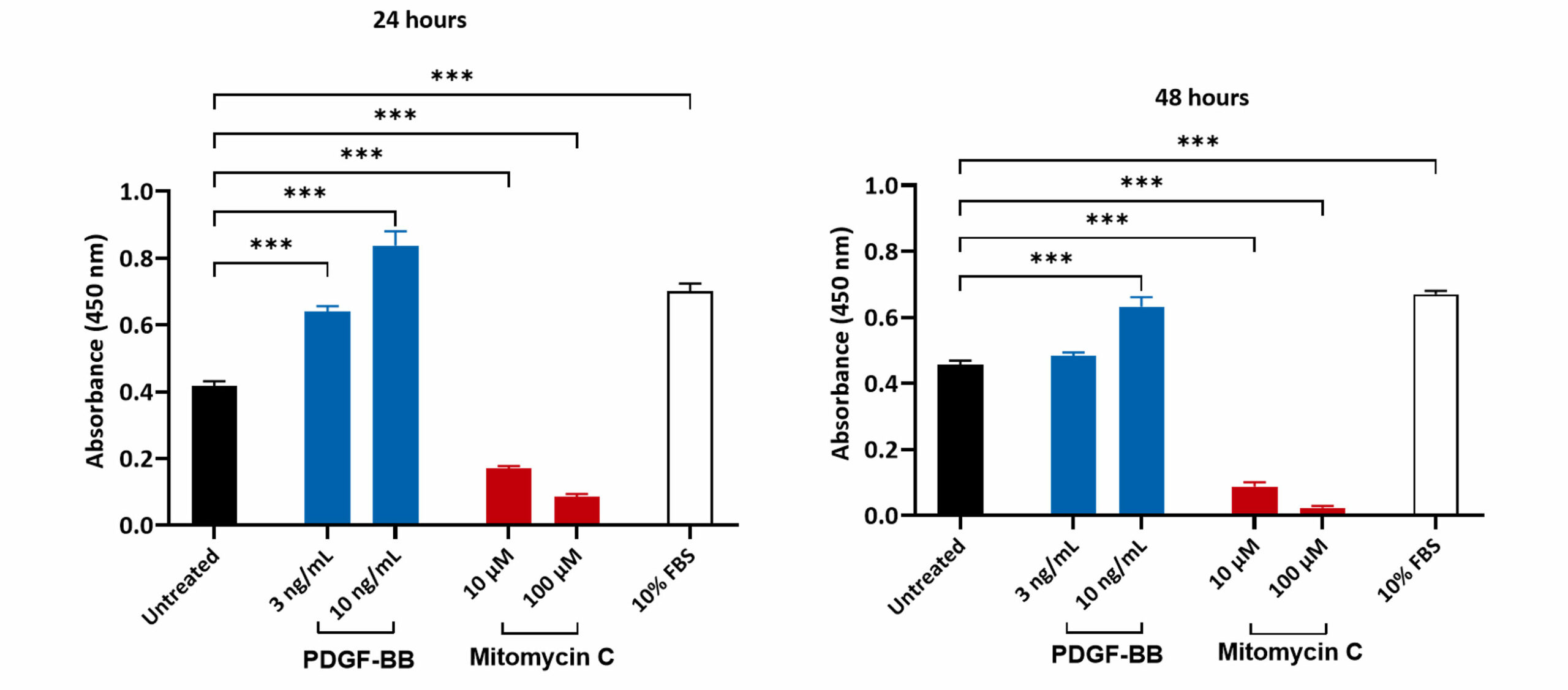
Bromodeoxyuridine (BrdU) assay to measure proliferation. HLF (human lung fibroblasts) cells were seeded at an optimised density and incubated overnight at 37oC in a humidified incubator. The cells were treated with PDGF-BB (to promote proliferation) and Mitomycin C (inhibits DNA synthesis/function). The BrdU assay showed that treatment with PDGF-BB and 10% FBS resulted in a significant increase in the cell proliferation, as indicated by the increase in the absorbance. Treatment with Mitomycin C significantly reduced the absorbance, suggesting an inhibition in cell proliferation (***p<0.001 ± SEM).
MTT
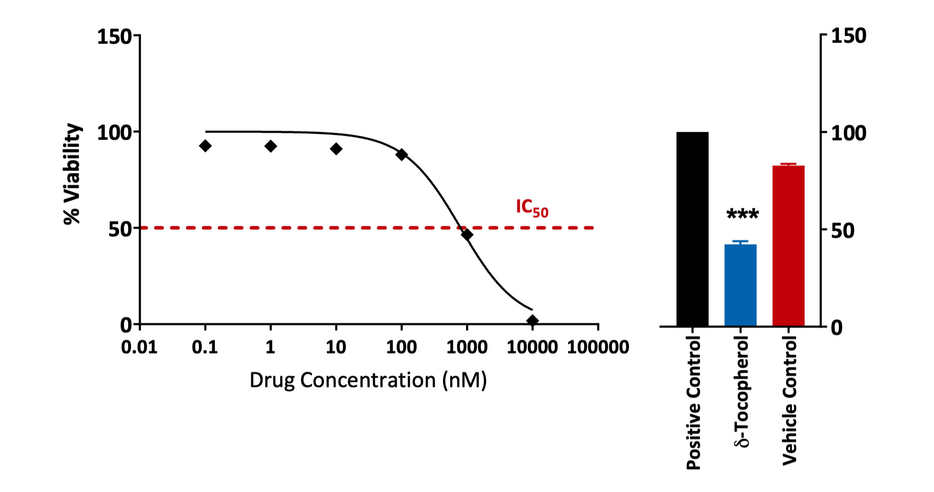
MTT assay to measure proliferation. HMC1.2 cells were treated with a test compound at 6 different concentrations. The % cell viability post-compound-treatment was determined by means of a MTT assay (assesses metabolically active live cells). A significant reduction in cell viability was observed with δ-Tocopherol when compared to the positive and vehicle controls (***p<0.001; n=5; ±SEM)
Alamar Blue
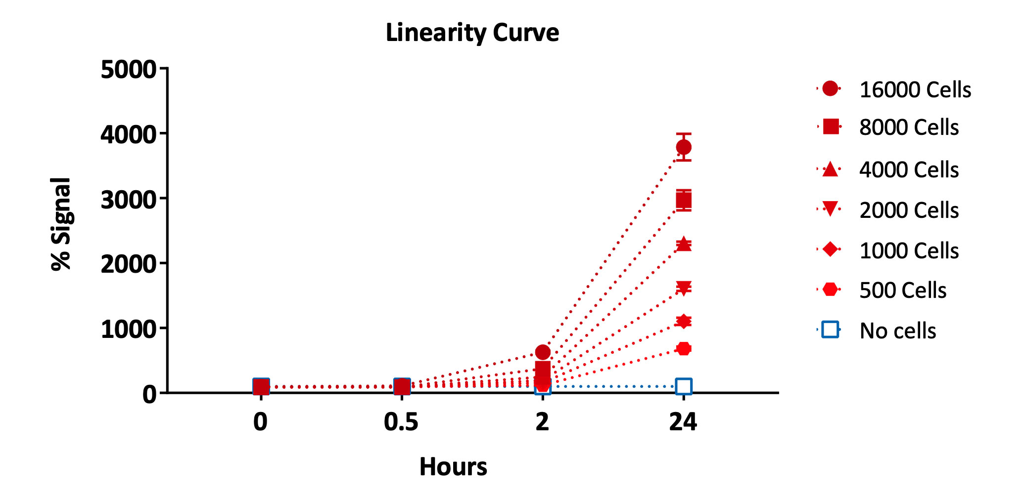
Metabolic activity assay (Alamar blue™) of Human umbilical vein endotheial cells (HUVEC). Cells were seeded at different densities and the proliferation was recorded over 24 hours using Alamar Blue dye as % signal (signal increases in the reducing environment of viable cells).
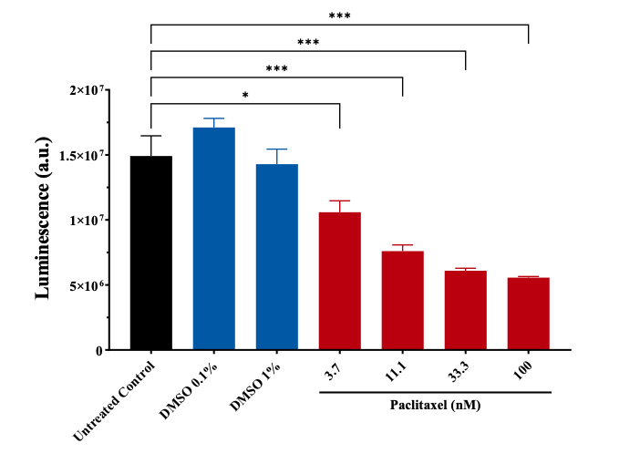
Metabolic activity assay (Alamar blue™) of NCI-H460 cells treated with Paclitaxel. Data presented as mean ± SEM of fluorescence signal, performed in technical triplicate. Statistical significance determined by Ordinary one-way ANOVA followed by Dunnett’s T-test for correction of multiple comparisons (* p<0.033; ** p<0.002; *** p<0.001).
Request a consultation with Cellomatics Biosciences today
Our experienced team of in vitro laboratory scientists will work with you to understand your project and provide a bespoke project plan with a professional, flexible service and a fast turnaround time.
To request a consultation where we can discuss your exact requirements, please contact Cellomatics Biosciences.


