


Peripheral blood mononuclear cells (PBMC) provide selective responses to the immune system and are the major immune cells in the human body. PBMCs include lymphocytes (T cells, B cells, and NK cells), monocytes, and dendritic cells. PBMC based assays explore the role of these cells in immune modulation, cell proliferation/cytotoxicity and their role in targeting specific cancer cells.
The expert team at Cellomatics can support with PBMC assays within a variety of projects.
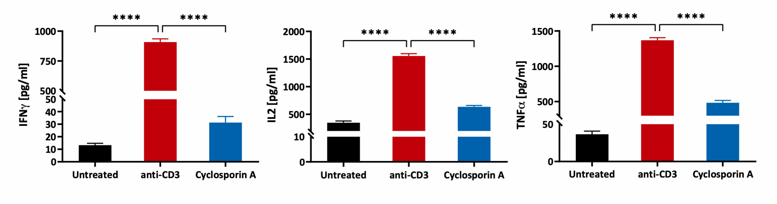
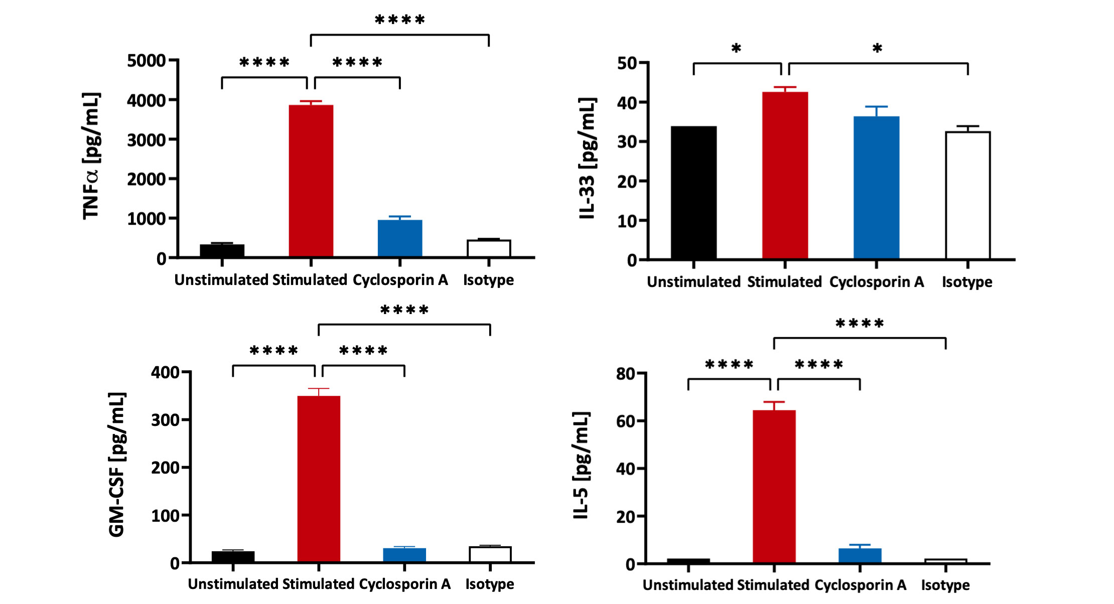
Freshly isolated Peripheral Blood Mononuclear cells (PBMCs) were seeded onto anti-CD3 coated U-bottom plates and further stimulated with soluble anti-CD28 for 48 hours. PBMCs were also treated with Cyclosporin A prior to stimulation. Isotype of anti-CD3 was used as a background control. Supernatants were analysed for inflammatory markers using a Luminex Multiplex Assay (n=3±SEM; *p<0.05; ****p<0.0001).
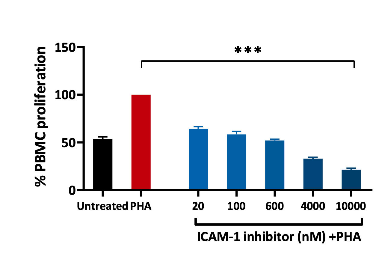
Effect of ICAM-1 inhibitor on PBMC proliferation.
PHA induced an increase in PBMC proliferation. A significant reduction in cell proliferation was observed when cells were pre-treated with ICAM-1 inhibitor. Statistical analysis performed using a one-way ANOVA (*p<0.05; **p<0.01; ***p<0.001; ±SEM).
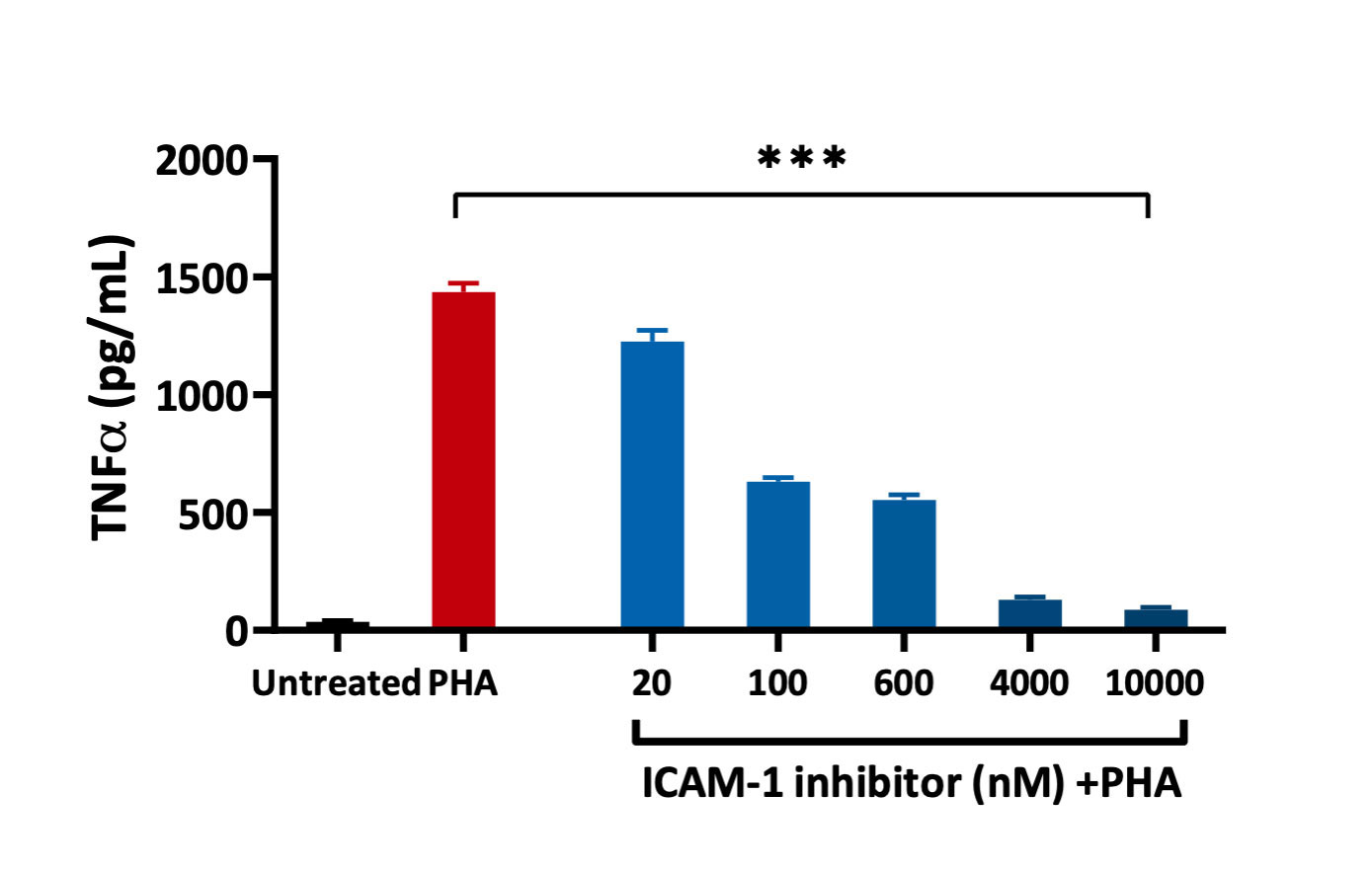
Effect of ICAM-1 inhibitor on TNFα release
PHA stimulation of PBMCs significantly increased the expression level of TNFα. A dose-dependent reduction was observed for cells treated with ICAM-1 inhibitor. Statistical analysis performed using a one-way ANOVA (**p<0.01; ***p<0.001; ±SEM).
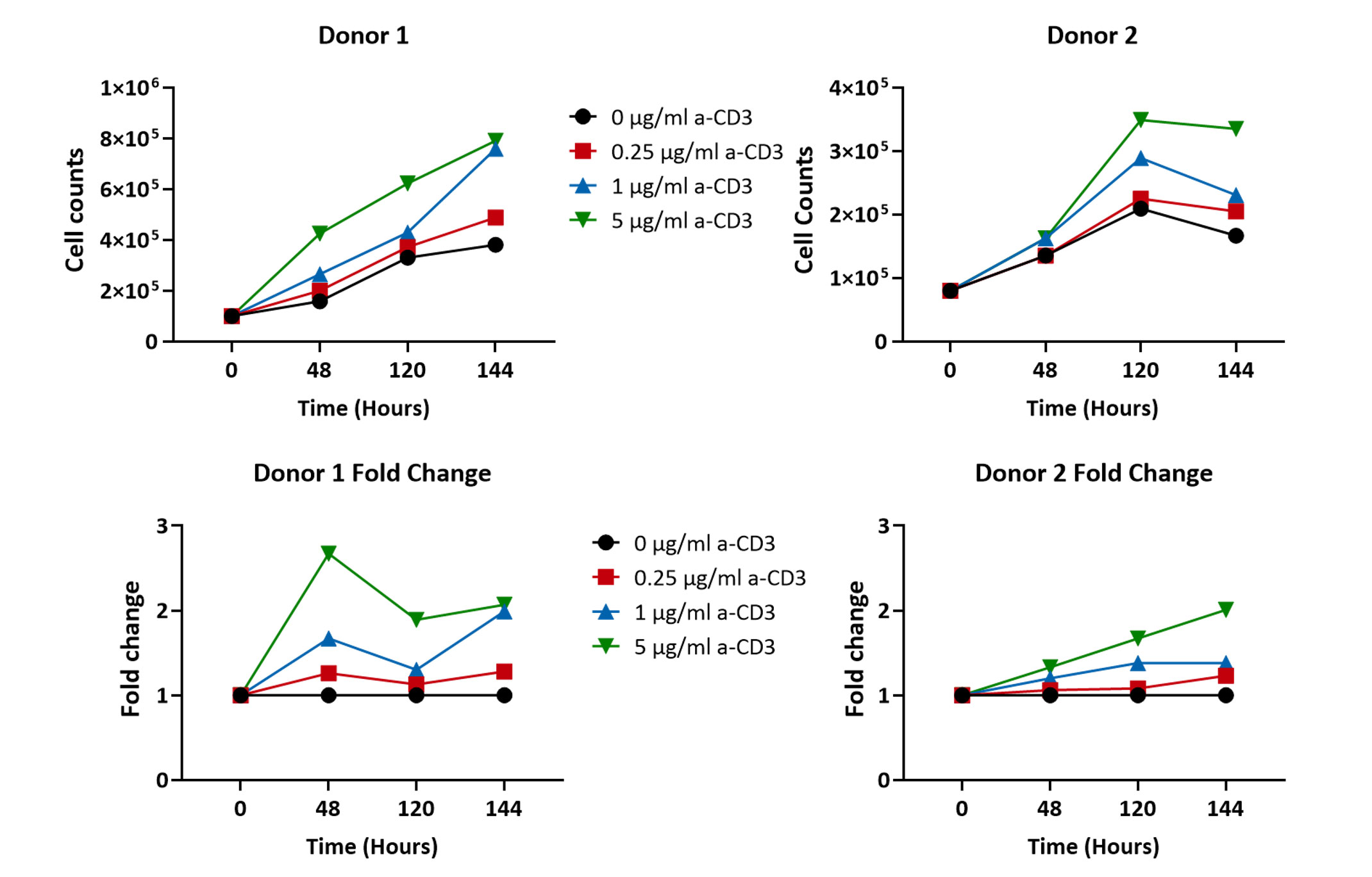
PBMCs from 2 different healthy volunteers were stimulated with increasing concentrations of anti-CD3 antibody (0 µg/ml, 0.25 µg/ml, 1 µg/ml and 5 µg/ml) over the period of 144 hours (6 days) and the cell proliferation was recorded. A concentration dependent increase in PBMC cell counts was observed after 48 hours of stimulation (n=5±SEM).
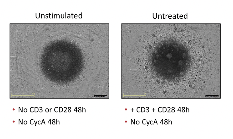
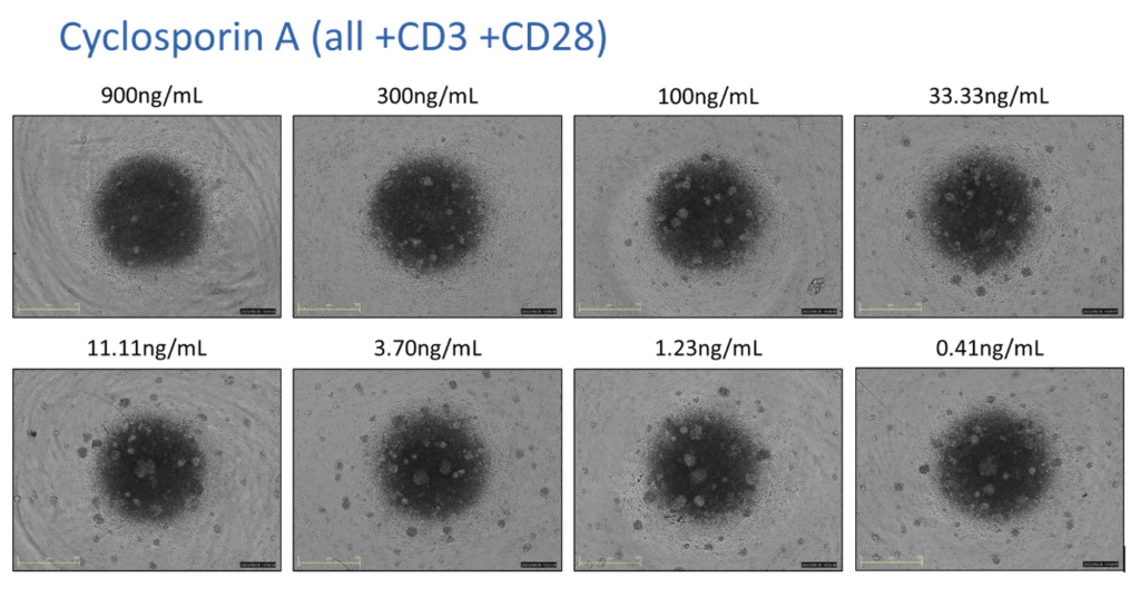
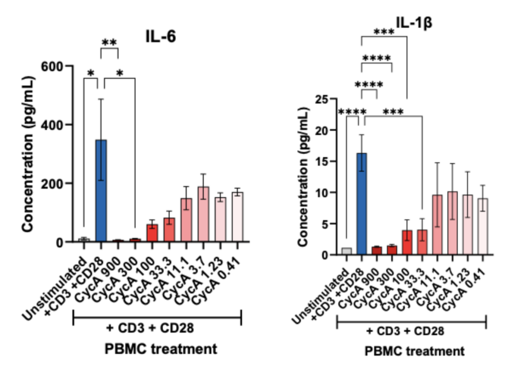
PBMCs – anti-CD3/anti-CD28 stimulation Freshly isolated Peripheral Blood Mononuclear cells (PBMCs) were seeded onto anti-CD3 coated U-bottom plates and further stimulated with soluble anti-CD28 for 48 hours. PBMCs were also treated with Cyclosporin A in a 8 concentration-response-curve prior to stimulation. Supernatants were analysed for inflammatory markers using Luminex Multiplex Assay (n=3±SEM; *p<0.05; ****p<0.0001).
PBMCs – anti-CD3/anti-CD28 stimulation Freshly isolated Peripheral Blood Mononuclear cells (PBMCs) were seeded onto anti-CD3 coated U-bottom plates and further stimulated with soluble anti-CD28 for 48 hours. PBMCs were also treated with Cyclosporin A prior to stimulation. Isotype of anti-CD3 was used as a background control. Supernatants were analysed for inflammatory markers using Luminex Multiplex Assay (n=3±SEM; *p<0.05; ****p<0.0001
Freshly isolated Peripheral Blood Mononuclear cells (PBMCs) were seeded onto anti-CD3 coated U-bottom plates and further stimulated with soluble anti-CD28 for 24, 48 and 72 hours. PBMCs were also treated with Cyclosporin prior to stimulation. Isotype of anti-CD3 was used as a background control. Supernatants were analysed for inflammatory markers using Luminex Multiplex Assay (n=5±SEM; **p<0.01; ***p<0.001; ****p<0.0001).
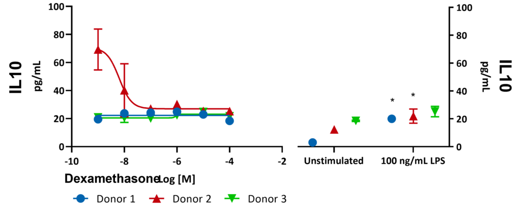
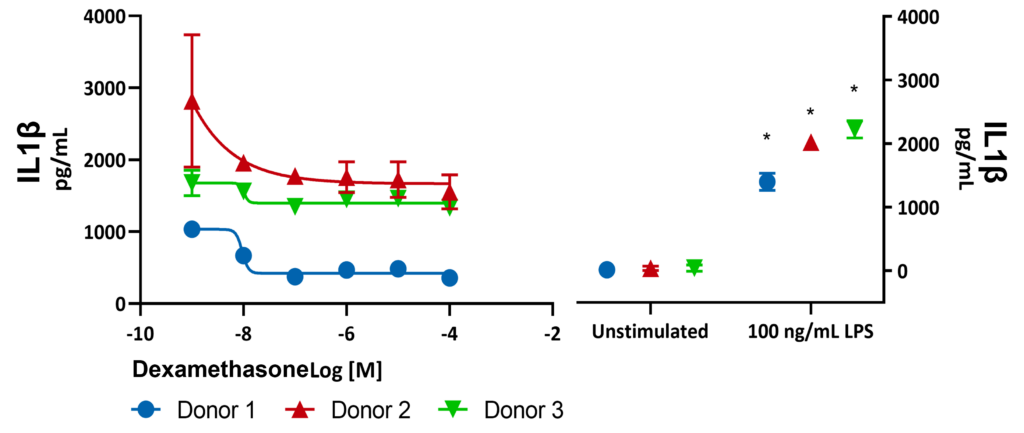
Freshly isolated Peripheral Blood Mononuclear cells (PBMCs) were seeded onto anti-CD3 coated U-bottom plates and further stimulated with LPS for 48 hours in the presence or absence of Dexamethasone. Supernatants were analysed for inflammatory markers using a Luminex Multiplex Assay (n=3±SEM; *p<0.05; ****p<0.0001).
Freshly isolated Peripheral Blood Mononuclear cells (PBMCs) from 3 donors were stimulated with LPS for 24 hours in the presence or absence of Dexamethasone. Supernatants were analysed for inflammatory markers using Luminex Multiplex Assay. There was a significant increase in TNF-α expression with 100 ng/mL LPS (***p<0.001). Significant differences were identified using a two-way ANOVA followed by a Šídák’s multiple comparisons test.
Freshly isolated Peripheral Blood Mononuclear cells (PBMCs) from 3 donors were stimulated with LPS for 24 hours in the presence or absence of Dexamethasone. Supernatants were analysed for inflammatory markers using Luminex Multiplex Assay. There was a significant increase in IL6 expression with 100 ng/mL LPS (***p<0.001). Significant differences were identified using a two-way ANOVA followed by a Šídák’s multiple comparisons test.
Freshly isolated Peripheral Blood Mononuclear cells (PBMCs) from 3 donors were stimulated with LPS for 24 hours in the presence or absence of Dexamethasone. Supernatants were analysed for inflammatory markers using Luminex Multiplex Assay. There was a significant increase in IL10 expression with 100 ng/mL LPS (***p<0.001). Significant differences were identified using a two-way ANOVA followed by a Šídák’s multiple comparisons test.
Freshly isolated Peripheral Blood Mononuclear cells (PBMCs) from 3 donors were stimulated with LPS for 24 hours in the presence or absence of Dexamethasone. Supernatants were analysed for inflammatory markers using Luminex Multiplex Assay. There was a significant increase in IL1β expression with 100 ng/mL LPS (***p<0.001). Significant differences were identified using a two-way ANOVA followed by a Šídák’s multiple comparisons test.
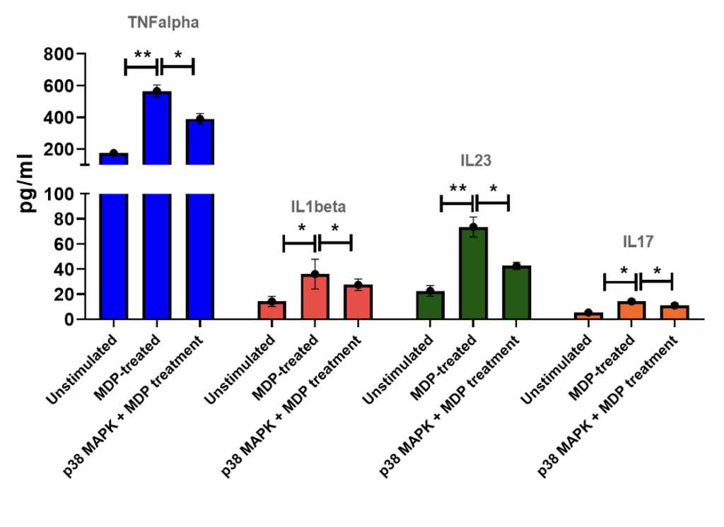
PBMCs + MDP (muramyl dipeptide) stimulation Levels of TNF alpha, IL1beta, IL-23 and IL-17 in PBMC supernatants of healthy volunteers following stimulation with MDP for 24 hrs in the absence or presence of p38MAPK inhibitor. Results are expressed in pg/mL.
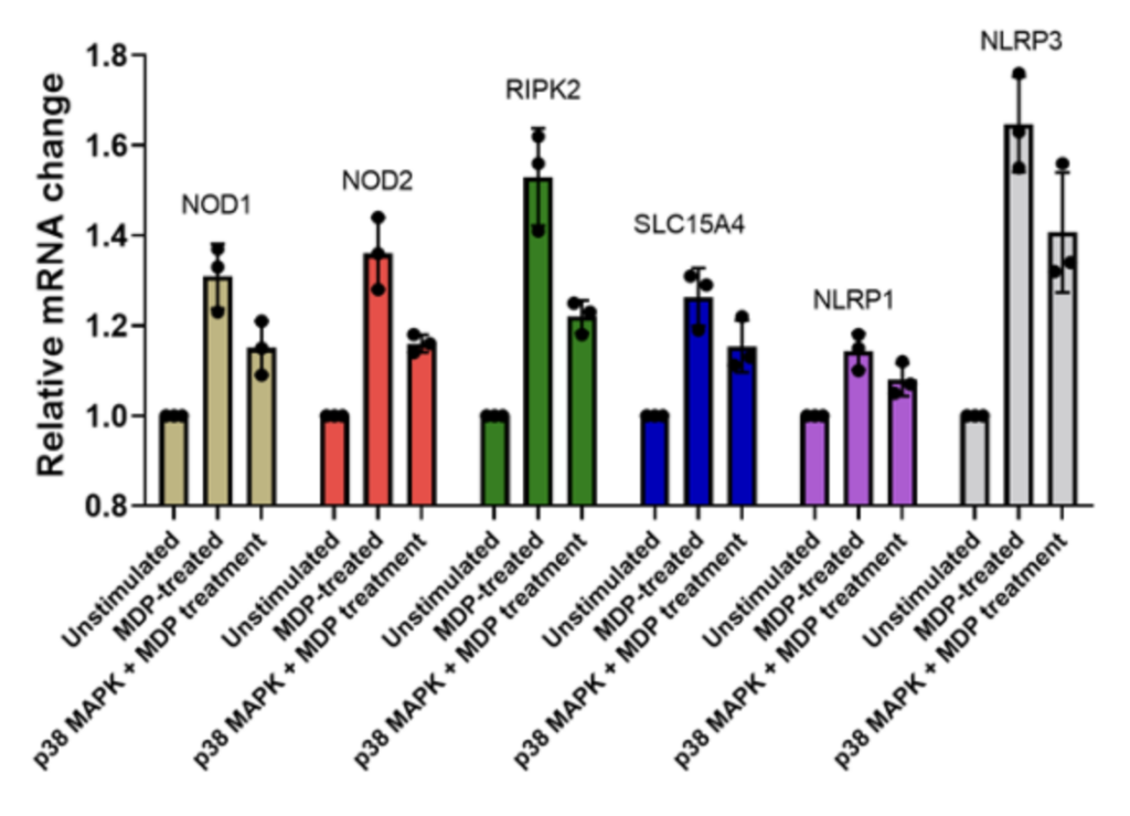
PBMCs + MDP (muramyl dipeptide) stimulation Relative mRNA levels of NOD1, NOD2, RIPK2, SLC15A4, NLRP1 and NLRP3 following stimulation of PBMCs (from a healthy volunteer) with MDP in the absence or presence of p38MAPK inhibitor.
PBMCs from healthy domors were stimulated with LPS alone, LPS+ATP in the presence or absence of NLRP3 inhibitor. Supernatants were analysed for NLRP3 and IL-1β using ELISA. Statistical analysis was performed using a 2WAY ANOVA, followed by Šídák’s multiple comparisons test for each donor independently. All treatment groups were compared to the untreated group (n=3 ± SEM; ***p<0.001, **p<0.01). Notably, the combination of LPS + ATP for 1 hr with 10µM NLRP3 inhibitor significantly reduced the expression of both NLRP3 and IL-1β in PBMCs from healthy donors.
PBMCs from healthy donors were stimulated with LPS alone, LPS+ATP in the presence or absence of NLRP3 inhibitor. Supernatants were analyzed using ELISA for NLRP3, IL-1β and Caspase 1. Data was subjected to statistical analysis using an ordinary one-way ANOVA, followed by a Dunnett’s multiple comparisons test for each donor independently. All treatment groups were compared to the LPS+ATP group (n=3 ± SEM; ***p<0.001, **p<0.01). The combination of 10µM-1µM NLRP3 inhibitor with LPS and ATP significantly reduced the expression of both NLRP3 and IL-1β in healthy PBMCs.
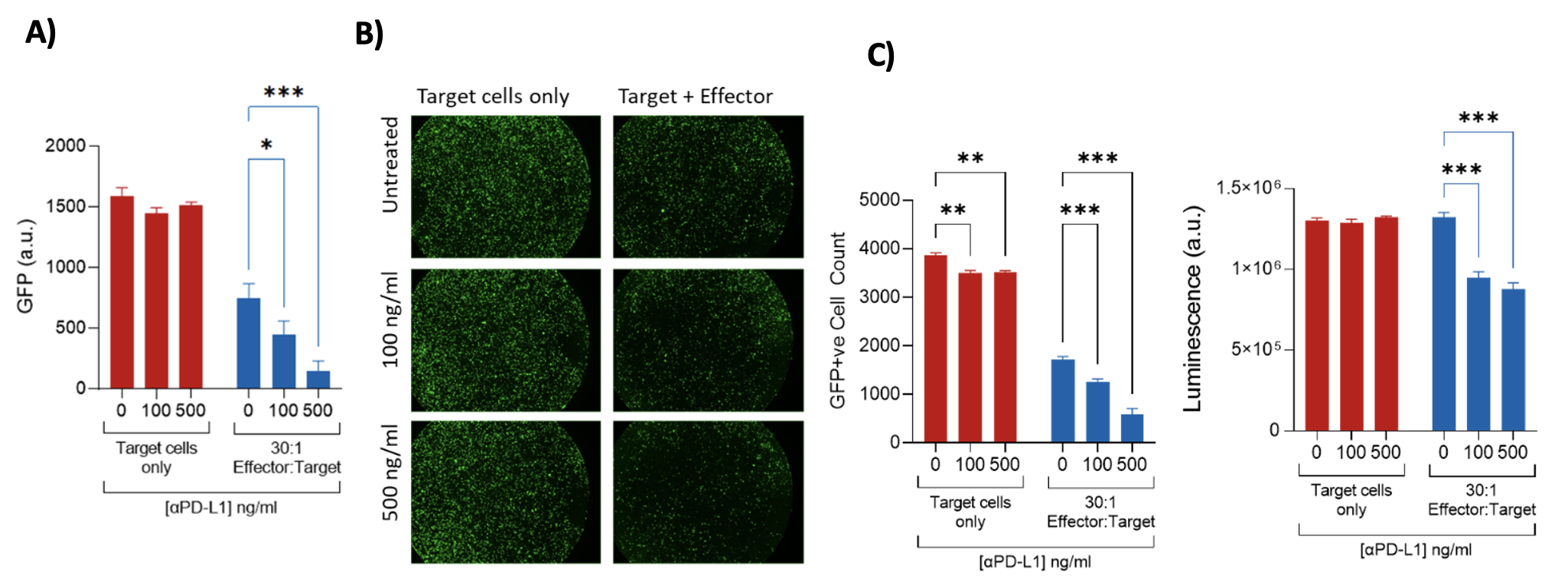
Assessment of ADCC via GFP release – MB231 (target cells)/PBMCs(effector cells) MB231 target cells were lipofected with a GFP expression plasmid and incubated with an antibody targeting human PD-L1-hlgG1fut. Target cells were subsequently incubated for 24 h with PBMCs (Effector cells) at a ratio of 30:1. ADCC was assessed by means of loss of intracellular GFP (GFP levels were measured on a plate reader, and the number of GFP+ve cells quantified by image analysis) and loss of viability (CellTiter Glo assay). A) A loss of intracellular GFP (demonstrating ADCC of GFP+ve Target cells) is induced by the Effector cells and is significantly increased by the antibody; B) Visualisation and subsequent image analysis demonstrate a loss of GFP+ve Target cells in the presence of the Effector cells, which is significantly enhanced by incubation with the antibody: C) A loss of viability occurs following incubation of Target cells with the antibody and Effector cells.
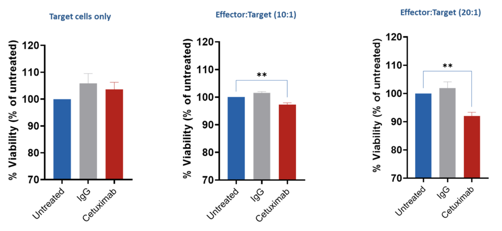
Assessment of ADCC vis GFP release – A549 (target cells)/PBMCs (effector cells) A549 (human lung adenocarcinoma) cells were co-cultured with PBMCs from a single donor for 4 hours in the presence or absence of Cetuximab (EGFR inhibitor antibody) for 4 hours to investigate the ADCC effect. CellTiterGlo assay (ATP-based viability) was performed, and results expressed as % of untreated cells.
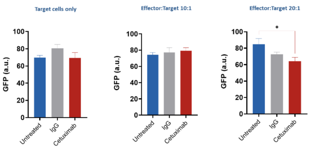
Assessment of ADCC via GFP release – HCT116 (target cells)/PBMCs(effector cells) HCT116 (human colorectal carcinoma cell) cells were co-cultured with PBMCs from a single donor for 4 hours in the presence or absence of Cetuximab (EGFR inhibitor antibody) for 4 hours to investigate the ADCC effect. Intracellular GFP levels was measured, and results expressed as relative fluorescent units.
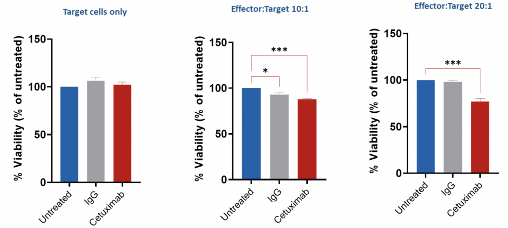
Assessment of ADCC via GFP release – HCT116 (target cells)/PBMCs(effector cells) HCT116 (human colorectal carcinoma cell) cells were co-cultured with PBMCs from a single donor for 4 hours in the presence or absence of Cetuximab (EGFR inhibitor antibody) for 4 hours to investigate the ADCC effect. CellTiterGlo assay (ATP-based viability) was performed, and results expressed as % of untreated cells.
Our experienced team of in vitro laboratory scientists will work with you to understand your project and provide a bespoke project plan with a professional, flexible service and a fast turnaround time.
To request a consultation where we can discuss your exact requirements, please contact Cellomatics Biosciences.
Cellomatics Biosciences Limited
10 Colwick Quays Business Park
Road No2, Colwick Nottingham NG4 2JY, UK
+44 (115) 865 4101
info@cellomaticsbio.com

Cellomatics Biosciences Limited
10 Colwick Quays Business Park
Road No2, Colwick Nottingham NG4 2JY, UK
+44 (115) 865 4101
info@cellomaticsbio.com


| Cookie | Duration | Description |
|---|---|---|
| cookielawinfo-checkbox-analytics | 11 months | This cookie is set by GDPR Cookie Consent plugin. The cookie is used to store the user consent for the cookies in the category "Analytics". |
| cookielawinfo-checkbox-functional | 11 months | The cookie is set by GDPR cookie consent to record the user consent for the cookies in the category "Functional". |
| cookielawinfo-checkbox-necessary | 11 months | This cookie is set by GDPR Cookie Consent plugin. The cookies is used to store the user consent for the cookies in the category "Necessary". |
| cookielawinfo-checkbox-others | 11 months | This cookie is set by GDPR Cookie Consent plugin. The cookie is used to store the user consent for the cookies in the category "Other. |
| cookielawinfo-checkbox-performance | 11 months | This cookie is set by GDPR Cookie Consent plugin. The cookie is used to store the user consent for the cookies in the category "Performance". |
| viewed_cookie_policy | 11 months | The cookie is set by the GDPR Cookie Consent plugin and is used to store whether or not user has consented to the use of cookies. It does not store any personal data. |