


Idiopathic pulmonary fibrosis (IPF) is the commonest interstitial lung disease (ILD) and is characterised by progressive scarring of the lungs. An impaired pulmonary wound healing response following lung injury coupled with excessive production of collagens leads to a pathogenic lung fibrosis.
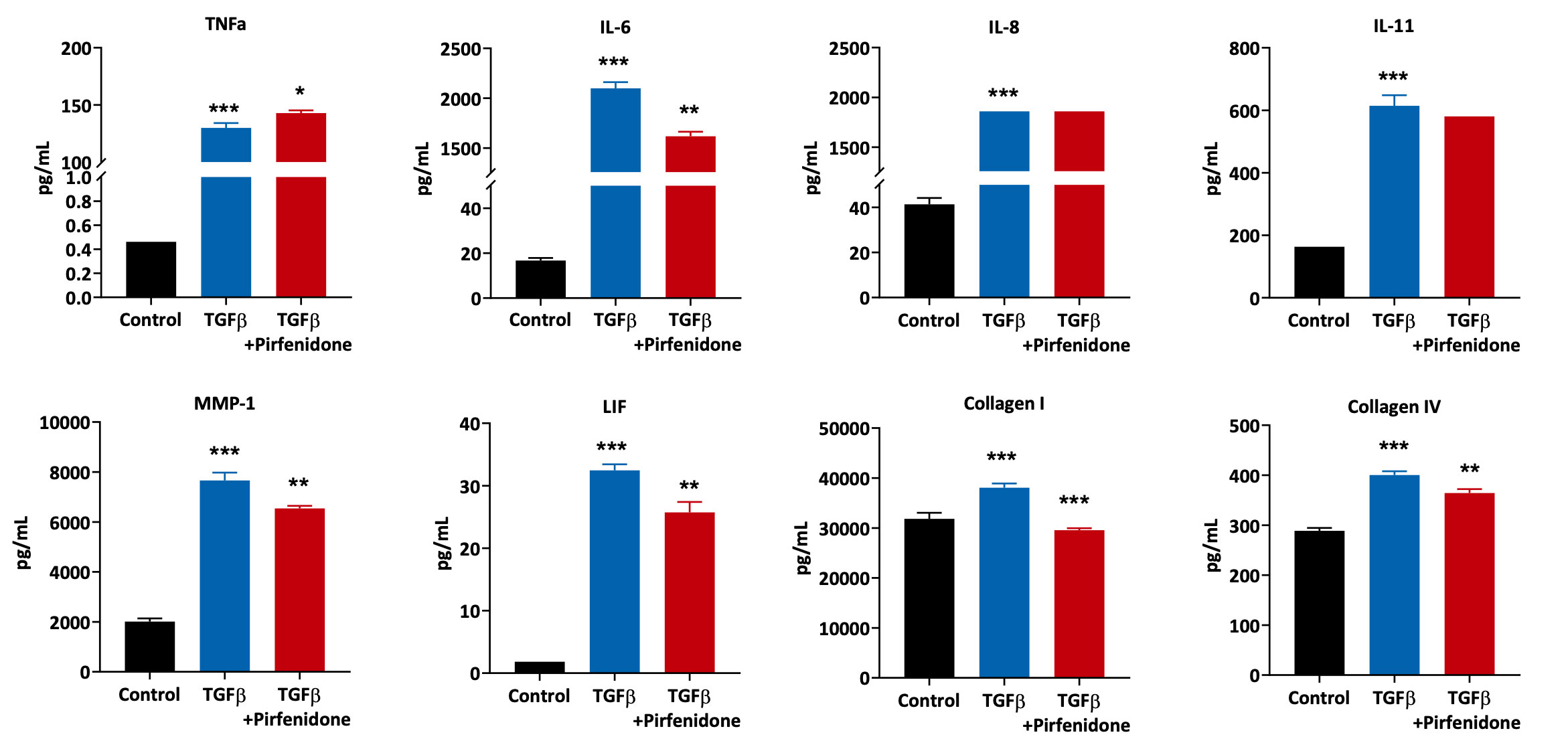
Primary Human Lung Fibroblasts were pre-treated with Pirfenidone for 1 hour followed by 24 hours stimulation with TGFβ. Supernatants were analysed using Luminex multiplexed assays for inflammatory and extracellular matrix component markers. All treatment conditions were compared to the TGFβ treated samples (***p<0.001; **p<0.01; *p<0.05; n=3±SEM).
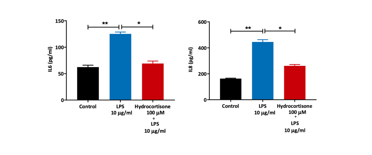
Primary Human Lung Fibroblasts were treated with 10 µg/ml LPS for 24 hours in the presence or absence of reference compound (hydrocortisone). Cell culture supernatants were then collected and levels of the proinflammatory IL6 and IL8 measured by ELISA (n=5±SEM; *p<0.05; **p<0.01).
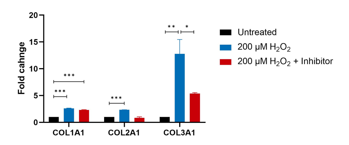
Primary Human Lung Fibroblasts were pre-treated with Pirfenidone (Targeting the inflammasome and down regulating TGFβ) for 1 hour followed by 24 hours stimulation with H2O2. Cell lysates were analysed using QuantiGene multiplex to detect changes in Collagen genes. Fold changes were determined by normalising target gene expression to house keeping genes (GAPDH and HPRT1). Treatment conditions were compared to the H2O2 treated samples (***p<0.001; **p<0.01; *p<0.05; n=3±SEM).
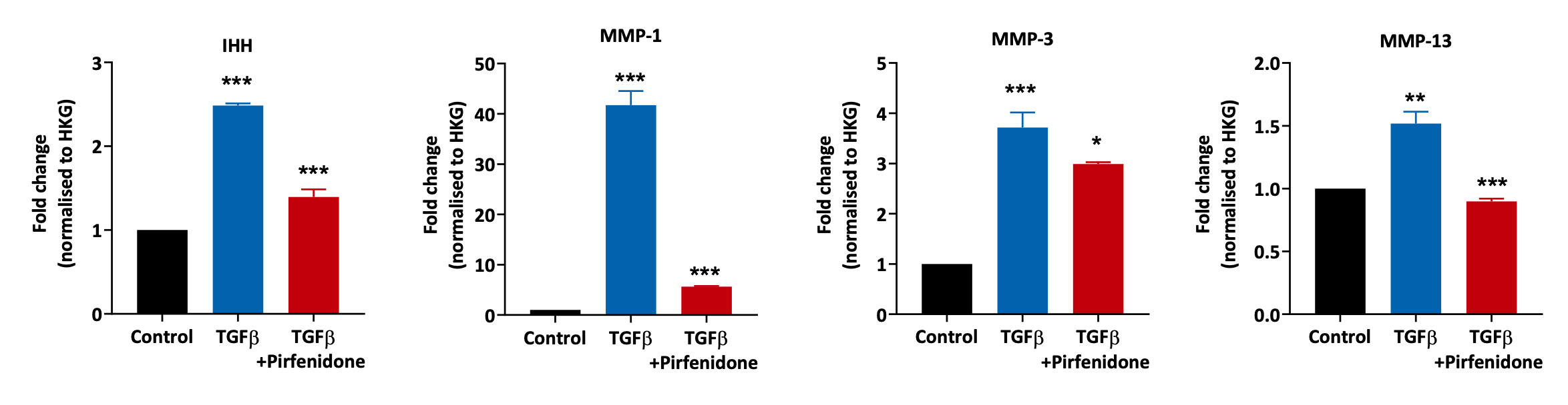
Primary Human Lung Fibroblasts were pre-treated with Pirfenidone for 1 hour followed by 24 hours stimulation with TGFβ (to drive the fibrotic pathway). Cell lysates were analysed using QuantiGene multiplex to detect changes in extracellular matrix component genes. Fold changes were determined by normalising target gene expression to house keeping genes (GAPDH and HPRT1). All treatment conditions were compared to the TGFβ treated samples (***p<0.001; **p<0.01; *p<0.05; n=3±SEM).
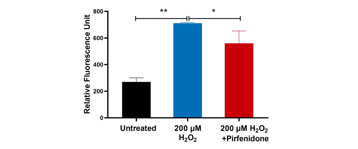
Human Lung Fibroblasts were pre-treated with Pirfenidone as an inhibitor for 1 hour followed by treatment with 200 µM H2O2 for 48 hours. Lysates were analysed for intracellular Reactive Oxygen Species (ROS) using a fluorescence detection kit. A significant increase in intracellular ROS (indicated by increased fluorescence signal) was observed in 200 µM H2O2 when compared to untreated controls. (n=3±SEM; **p<0.01).
Our experienced team of in vitro laboratory scientists will work with you to understand your project and provide a bespoke project plan with a professional, flexible service and a fast turnaround time.
To request a consultation where we can discuss your exact requirements, please contact Cellomatics Biosciences.
Cellomatics Biosciences Limited
10 Colwick Quays Business Park
Road No2, Colwick Nottingham NG4 2JY, UK
+44 (115) 865 4101
info@cellomaticsbio.com


Cellomatics Biosciences Limited
10 Colwick Quays Business Park
Road No2, Colwick Nottingham NG4 2JY, UK
+44 (115) 865 4101
info@cellomaticsbio.com




| Cookie | Duration | Description |
|---|---|---|
| cookielawinfo-checkbox-analytics | 11 months | This cookie is set by GDPR Cookie Consent plugin. The cookie is used to store the user consent for the cookies in the category "Analytics". |
| cookielawinfo-checkbox-functional | 11 months | The cookie is set by GDPR cookie consent to record the user consent for the cookies in the category "Functional". |
| cookielawinfo-checkbox-necessary | 11 months | This cookie is set by GDPR Cookie Consent plugin. The cookies is used to store the user consent for the cookies in the category "Necessary". |
| cookielawinfo-checkbox-others | 11 months | This cookie is set by GDPR Cookie Consent plugin. The cookie is used to store the user consent for the cookies in the category "Other. |
| cookielawinfo-checkbox-performance | 11 months | This cookie is set by GDPR Cookie Consent plugin. The cookie is used to store the user consent for the cookies in the category "Performance". |
| viewed_cookie_policy | 11 months | The cookie is set by the GDPR Cookie Consent plugin and is used to store whether or not user has consented to the use of cookies. It does not store any personal data. |