Atopic dermatitis (AD) is a chronic, complex allergic inflammatory skin disease that affects 10-20% of the population, characterised by a periodic flare of the symptoms which include red and itchy skin, swollen and cracked. Initial immune response begins with the T helper (Th)2 response which then shifts to Th1 in the chronic phase.
Our team have the expertise to offer multiple cell-based approaches including:
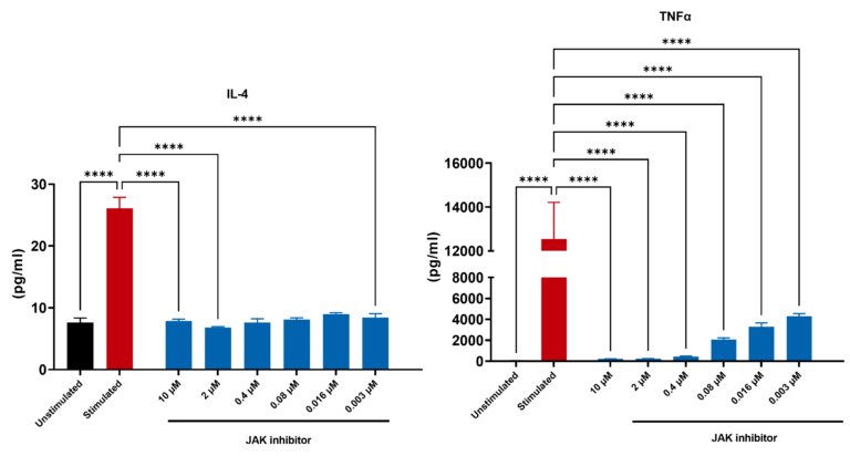
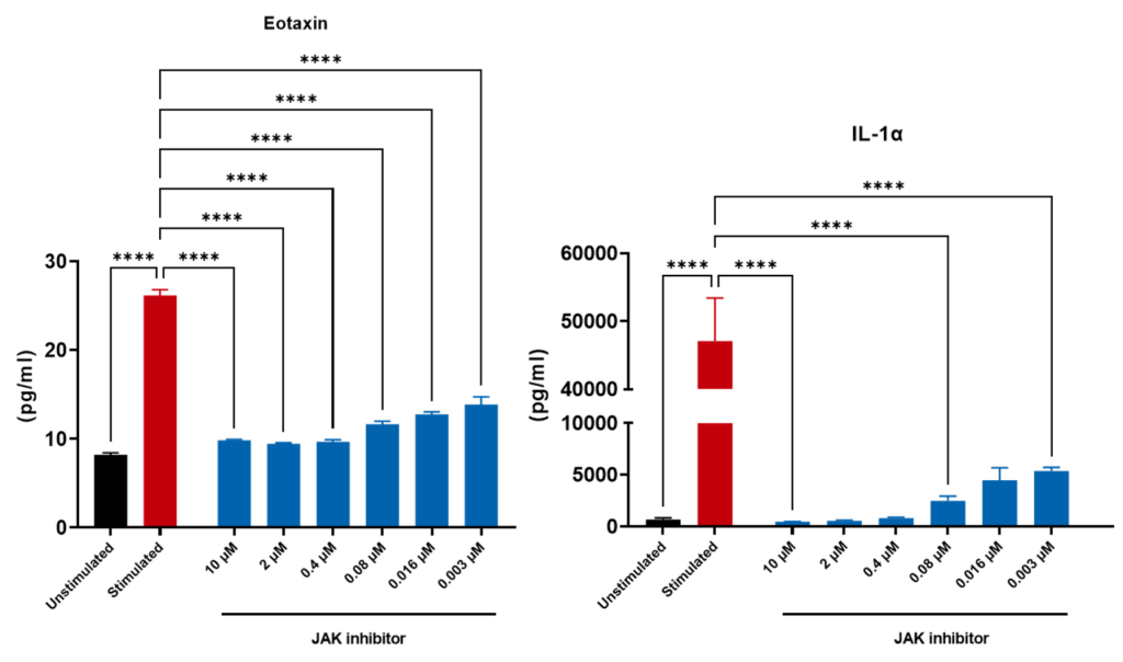
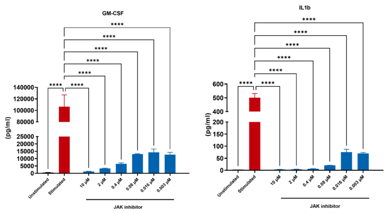
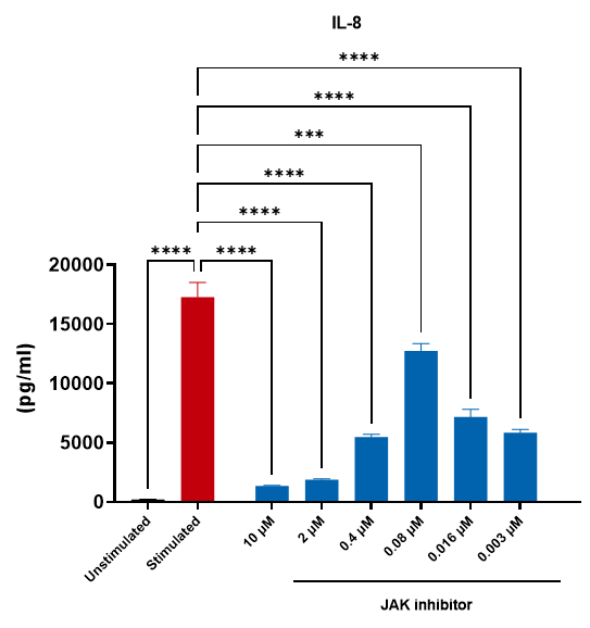
Atopic dermatitis in vitro model
Primary healthy human keratinocytes were treated with different concentrations of JAK inhibitor along with Poly I:C+ recombinant IL-13 for 48 hours. Inflammatory markers in the resulting supernatants were analysed using a Human Magnetic Luminex® Assay (n=3, mean±SEM)
Our experienced team of in vitro laboratory scientists will work with you to understand your project and provide a bespoke project plan with a professional, flexible service and a fast turnaround time.
To request a consultation where we can discuss your exact requirements, please contact Cellomatics Biosciences.
Cellomatics Biosciences Limited
10 Colwick Quays Business Park
Road No2, Colwick Nottingham NG4 2JY, UK
+44 (115) 865 4101
info@cellomaticsbio.com

Cellomatics Biosciences Limited
10 Colwick Quays Business Park
Road No2, Colwick Nottingham NG4 2JY, UK
+44 (115) 865 4101
info@cellomaticsbio.com


| Cookie | Duration | Description |
|---|---|---|
| cookielawinfo-checkbox-analytics | 11 months | This cookie is set by GDPR Cookie Consent plugin. The cookie is used to store the user consent for the cookies in the category "Analytics". |
| cookielawinfo-checkbox-functional | 11 months | The cookie is set by GDPR cookie consent to record the user consent for the cookies in the category "Functional". |
| cookielawinfo-checkbox-necessary | 11 months | This cookie is set by GDPR Cookie Consent plugin. The cookies is used to store the user consent for the cookies in the category "Necessary". |
| cookielawinfo-checkbox-others | 11 months | This cookie is set by GDPR Cookie Consent plugin. The cookie is used to store the user consent for the cookies in the category "Other. |
| cookielawinfo-checkbox-performance | 11 months | This cookie is set by GDPR Cookie Consent plugin. The cookie is used to store the user consent for the cookies in the category "Performance". |
| viewed_cookie_policy | 11 months | The cookie is set by the GDPR Cookie Consent plugin and is used to store whether or not user has consented to the use of cookies. It does not store any personal data. |