This is an imaging technique to aid microscopic anatomy of biological tissues.
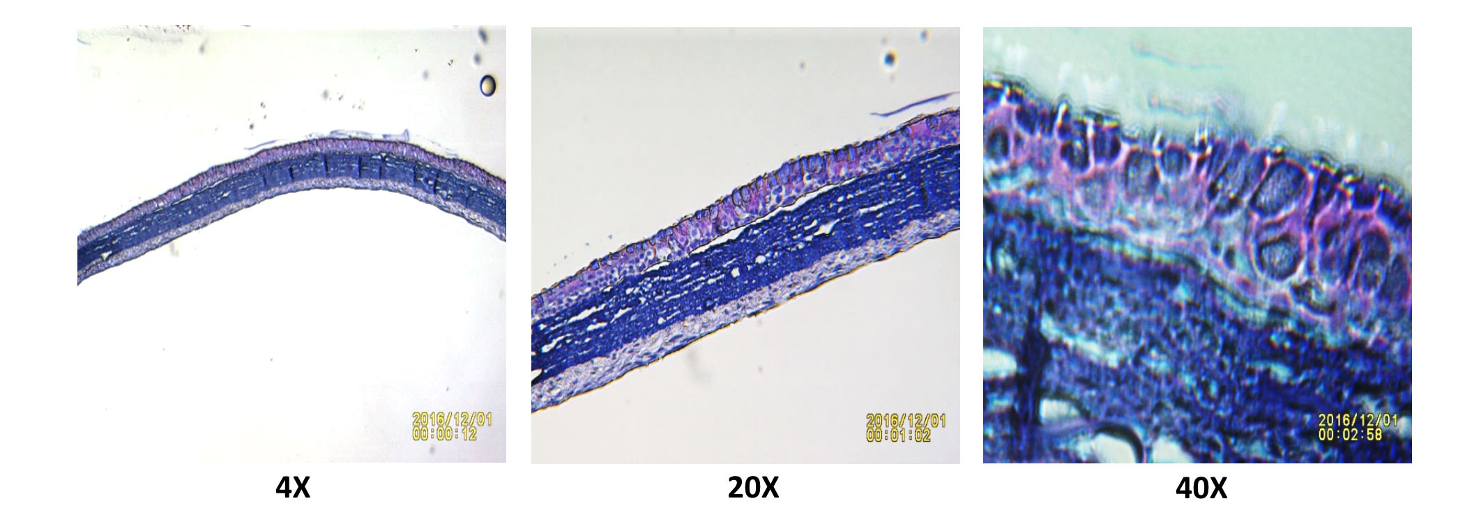
Normal Human Bronchial Epithelial Cells were grown on Air Liquid Interface (ALI) and allowed to differentiate for 28 days. At the end of the culture the cells were fixed and embedded into paraffin. The paraffin blocks were sectioned at 5µm thickness using Microtome and fixed onto slides. Haematoxylin and Eosin (H & E) staining were performed and slides were imaged at 4, 20 and 40X magnification. Distinct goblet and ciliated cells were observed on Day 28 of the ALI differentiation
Our experienced team of in vitro laboratory scientists will work with you to understand your phenotypic screening needs and provide a bespoke project plan with a professional, flexible service and a fast turnaround time.
To request a consultation where we can discuss your exact requirements, please contact Cellomatics Biosciences.
Cellomatics Biosciences Limited
10 Colwick Quays Business Park
Road No2, Colwick Nottingham NG4 2JY, UK
+44 (115) 865 4101
info@cellomaticsbio.com

Cellomatics Biosciences Limited
10 Colwick Quays Business Park
Road No2, Colwick Nottingham NG4 2JY, UK
+44 (115) 865 4101
info@cellomaticsbio.com


| Cookie | Duration | Description |
|---|---|---|
| cookielawinfo-checkbox-analytics | 11 months | This cookie is set by GDPR Cookie Consent plugin. The cookie is used to store the user consent for the cookies in the category "Analytics". |
| cookielawinfo-checkbox-functional | 11 months | The cookie is set by GDPR cookie consent to record the user consent for the cookies in the category "Functional". |
| cookielawinfo-checkbox-necessary | 11 months | This cookie is set by GDPR Cookie Consent plugin. The cookies is used to store the user consent for the cookies in the category "Necessary". |
| cookielawinfo-checkbox-others | 11 months | This cookie is set by GDPR Cookie Consent plugin. The cookie is used to store the user consent for the cookies in the category "Other. |
| cookielawinfo-checkbox-performance | 11 months | This cookie is set by GDPR Cookie Consent plugin. The cookie is used to store the user consent for the cookies in the category "Performance". |
| viewed_cookie_policy | 11 months | The cookie is set by the GDPR Cookie Consent plugin and is used to store whether or not user has consented to the use of cookies. It does not store any personal data. |