
Western blotting is a widely used analytical technique in molecular biology and immunogenetics to detect specific proteins in a sample of tissue homogenate or cell extracts.
For more information about how Cellomatics can support your project, contact us today.
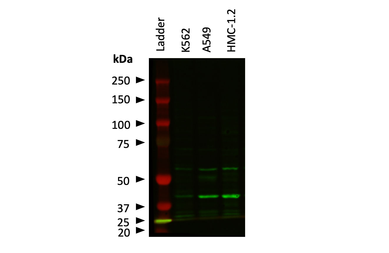
Near IR imaging of anti-RIP kinase protein conducted on cell lysates separated on a 8% SDS-PAGE gel.
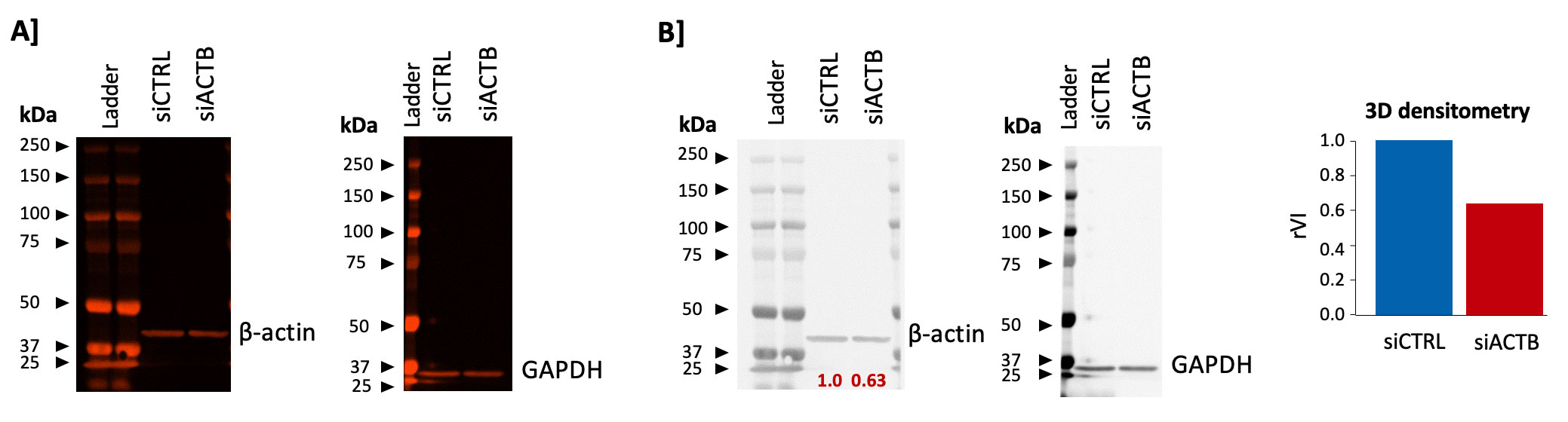
Human Primary Epithelial Cells were transfected with the following ON TARGETplus smart pool reagents: non-targeting control (siCTRL) and siACTB using Lipofectamine.
A] Near IR imaging of Human Primary Epithelial Cell lysates captured 24 hours post-transfection, after SDS-PAGE separation and immunoblotting using anti-β-actin and anti-GAPDH antibodies.
B] 3D densitometry calculated using the Q9 Alliance© Software, for figures reported in A]. Analysis confirmed reduction of β-actin band in cells transfected with the siACTB, when compared to controls (rVI=relative volume intensity).
Our experienced team of in vitro laboratory scientists will work with you to understand your cell-based assay development needs and provide a bespoke project plan with a professional, flexible service and a fast turnaround time.
To request a consultation where we can discuss your exact requirements, please contact Cellomatics Biosciences.
Cellomatics Biosciences Limited
10 Colwick Quays Business Park
Road No2, Colwick Nottingham NG4 2JY, UK
+44 (115) 865 4101
info@cellomaticsbio.com

Cellomatics Biosciences Limited
10 Colwick Quays Business Park
Road No2, Colwick Nottingham NG4 2JY, UK
+44 (115) 865 4101
info@cellomaticsbio.com


| Cookie | Duration | Description |
|---|---|---|
| cookielawinfo-checkbox-analytics | 11 months | This cookie is set by GDPR Cookie Consent plugin. The cookie is used to store the user consent for the cookies in the category "Analytics". |
| cookielawinfo-checkbox-functional | 11 months | The cookie is set by GDPR cookie consent to record the user consent for the cookies in the category "Functional". |
| cookielawinfo-checkbox-necessary | 11 months | This cookie is set by GDPR Cookie Consent plugin. The cookies is used to store the user consent for the cookies in the category "Necessary". |
| cookielawinfo-checkbox-others | 11 months | This cookie is set by GDPR Cookie Consent plugin. The cookie is used to store the user consent for the cookies in the category "Other. |
| cookielawinfo-checkbox-performance | 11 months | This cookie is set by GDPR Cookie Consent plugin. The cookie is used to store the user consent for the cookies in the category "Performance". |
| viewed_cookie_policy | 11 months | The cookie is set by the GDPR Cookie Consent plugin and is used to store whether or not user has consented to the use of cookies. It does not store any personal data. |