Invasion Assays
Invasion Assays
Cell invasion is a finely regulated processes that is critical in many normal physiological processes including during embryonic development, wound repair and immune surveillance (Lauffenburger and Horwitz, 1996; Pollard and Borisy, 2003; Ridley et al., 2003). However, these dynamic cell movements are also crucial in cancer progression and metastasis (Hamidi and Ivaska, 2018).
Here at Cellomatics, we support a wide range of projects with a focus on cell invasion (and migration) utilising well established invasion assays.
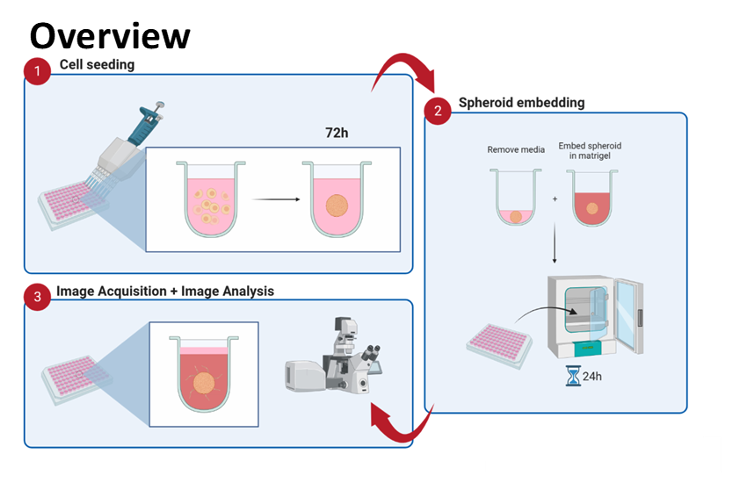
3D Spheroid Invasion Assays
Principle
- Plate method (based on self-assembling tumour spheroids) which has great potential for invasion studies.
- Semi-quantitative: microscopy and image analysis (Fiji/Image) calculate invaded area.
3D Spheroids and Invasion (HT-1080 cells - 24h)
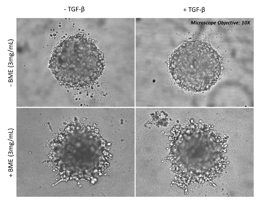
HT-1080 (human fibrosarcoma cell line) cells were grown as 3D spheroids for 72 hours before embedding in Basement Membrane Extract (BME) for 5-10 days in the presence or absence of TGFβ. Images were captured with a brightfield microscope – 10X
3D Spheroids and Invasion (NIH/3T3 cells - 24h)
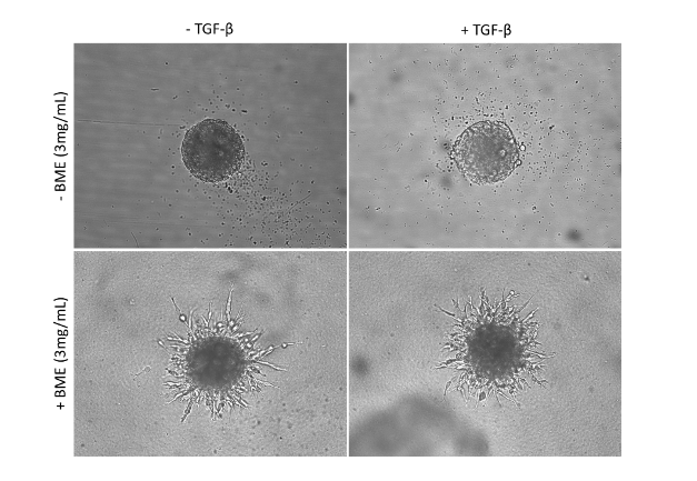
NIH/3T3 (mouse fibroblast cell line) cells were grown as 3D spheroids for 72 hours before embedding in Basement Membrane Extract (BME) for 5-10 days in the presence or absence of TGFβ. Images were captured with a brightfield microscope – 10X
3D Spheroids and Invasion (BEAS-2B and A549 cells)
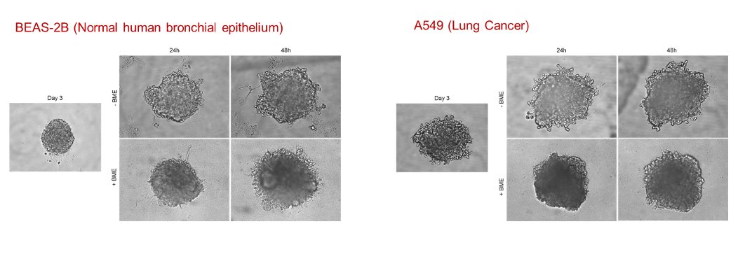
BEAS-2B (Normal human bronchial epithelium) and A549 (human lung epithelial cancer cell line) cells were grown as 3D spheroids for up to 72 hours with/without embedding in Basement Membrane Extract (BME) to study their invasive properties. Images were captured with a brightfield microscope – 10X
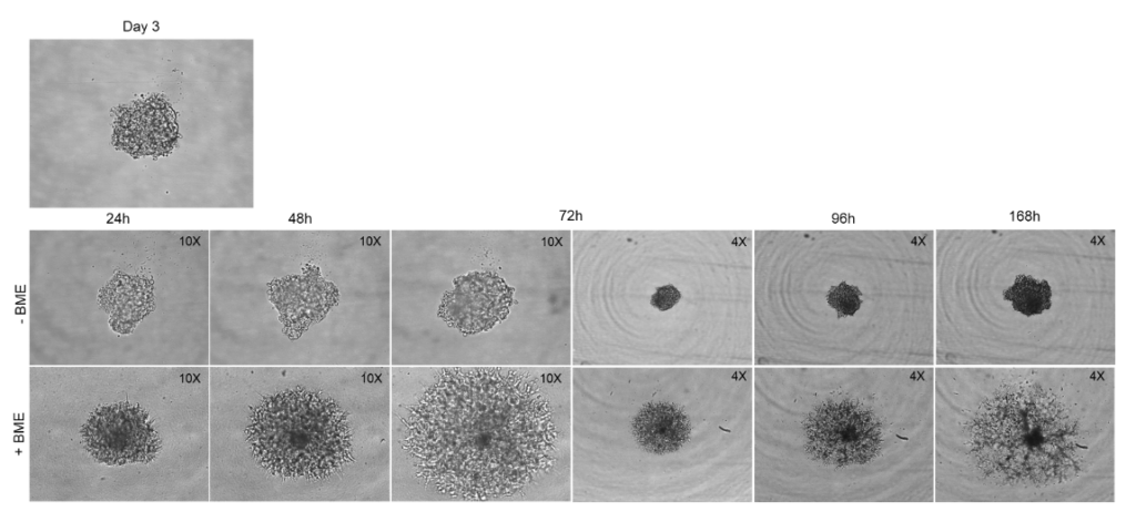
3D Spheroids and Invasion - SH-5YSY (Neuroblastoma)
SH-5YSY (Neuroblastoma) cells were grown as 3D spheroids for up to 7 days with/without embedding in Basement Membrane Extract (BME) to study their invasive properties. Images were captured with a brightfield microscope – 10X and 4X
3D Spheroid Invasion Assay - SH-5YSY (Neuroblastoma)
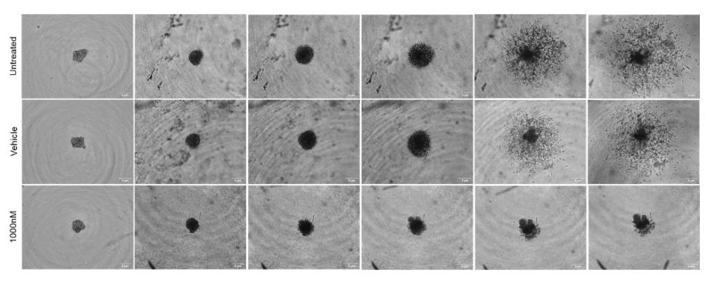
SH-5YSY (Neuroblastoma) cells were grown as 3D spheroids for up to 9 days by embedding in Basement Membrane Extract (BME) in the presence or absence of Cytochalasin D (potent inhibitor of actin polymerization). Images were captured with a brightfield microscope – 4X
3D Spheroid Invasion Assay - SH-5YSY (Neuroblastoma)
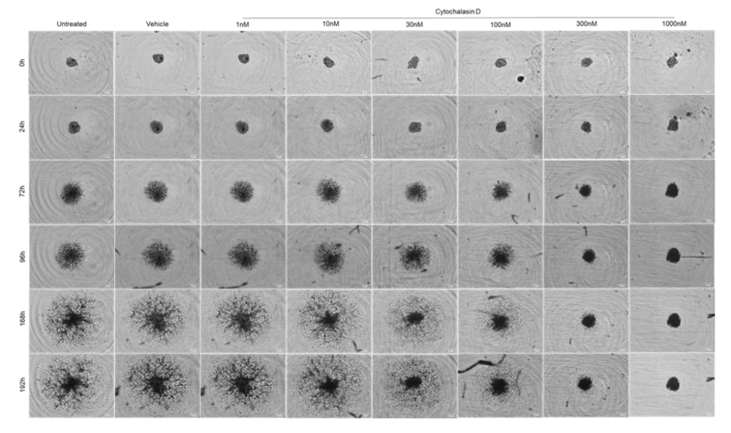
SH-5YSY (Neuroblastoma) cells were grown as 3D spheroids for up to 8 days by embedding in Basement Membrane Extract (BME) in the presence or absence of Cytochalasin D (potent inhibitor of actin polymerization) in a 6-concentration response curve. Images were captured with a brightfield microscope – 4X
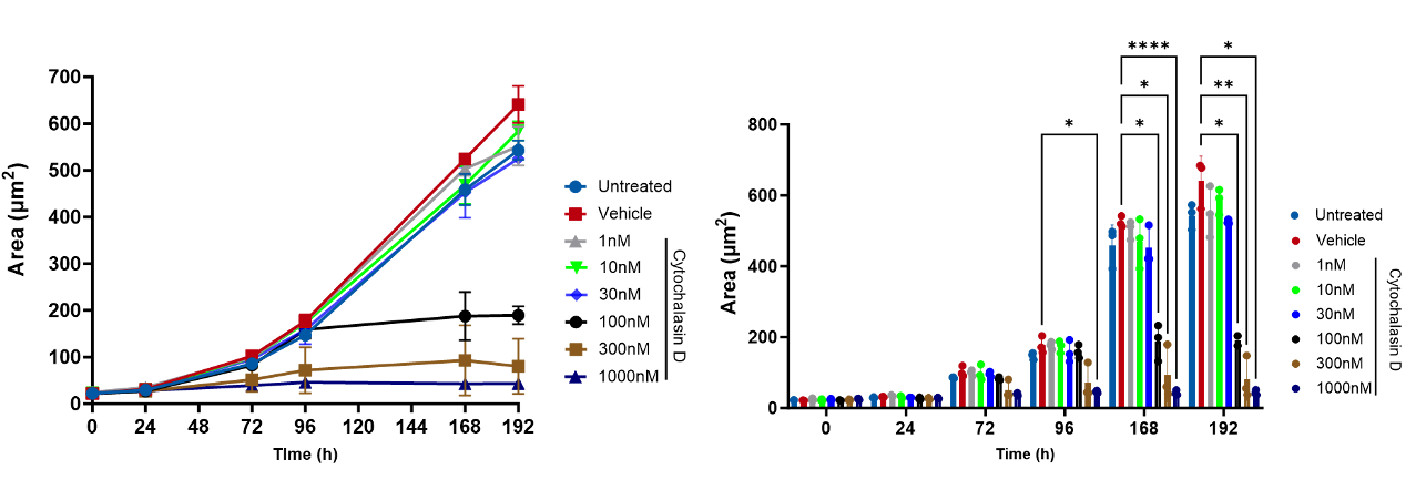
SH-5YSY (Neuroblastoma) cells were grown as 3D spheroids for up to 8 days by embedding in Basement Membrane Extract (BME) in the presence or absence of Cytochalasin D (potent inhibitor of actin polymerization) in a 6-concentration response curve. Images were captured with a brightfield microscope – 4X. Data analysed and platted as total spheroid area (µm2)
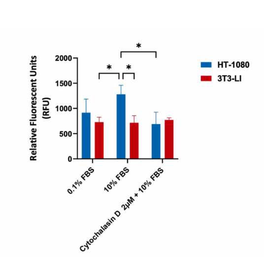
Invasion assay in Human Fibrosarcoma HT-1080 and mouse fibroblast 3T3-LI cells (negative control). Cells were seeded into Corning Transwell™ inserts and were coated with Cultrex® Basement Membrane Extract. Cells seeded in serum-free media invaded towards FBS for 48hrs in the presence or absence of Cytochalsain D. Invasion assay showed that treatment with 2µM Cytochalsain D resulted in a significant decrease in invasion with highly invasive HT-1080 cells, as indicated in the relative fluorescent values (fluorescence enhancement when bound to cellular nucleic acids; more cells more fluoresence).
Request a consultation with Cellomatics Biosciences today
Our experienced team of in vitro laboratory scientists will work with you to understand your project and provide a bespoke project plan with a professional, flexible service and a fast turnaround time.
To request a consultation where we can discuss your exact requirements, please contact Cellomatics Biosciences.


