


Pharmacology Services
GPCR Pharmacology Capabilities
At Cellomatics Biosciences, we provide specialist GPCR cell-based assay services and GPCR pharmacology contract research to support compound screening, pharmacological profiling, target validation and mechanism-of-action studies.
Our expertise in human cell-based in vitro models enables us to investigate GPCR-mediated signalling pathways, receptor activation and downstream cellular responses. Through customised assay development and functional pharmacological testing, we help pharmaceutical and biotechnology companies evaluate compound potency, characterise receptor interactions and generate reliable data to support drug discovery and preclinical development programmes.
Our GPCR Pharmacology capabilities
As a specialist GPCR functional pharmacology CRO, we take a flexible and bespoke approach, developing assay systems around the biological characteristics of the receptor, signalling pathway and compounds under investigation. Our capabilities include custom GPCR assay development, bespoke GPCR screening assays, GPCR signalling assays, GPCR pathway analysis and comprehensive GPCR compound characterisation.
For recombinant receptor studies, we can generate in-house polyclonal cell populations stably overexpressing GPCRs of interest and establish functional assays appropriate to the receptor’s downstream signalling pathway. Using second-messenger assays, including cAMP and IP-One measurements, we can interrogate GPCRs signalling through Gs, Gi and Gq pathways and determine key pharmacological parameters such as potency and efficacy. These robust and reproducible assay platforms can support concentration-response profiling, hit identification and the functional characterisation of agonists, antagonists and other GPCR modulators.
In addition to recombinant systems, our expertise in human cell-based GPCR assays and primary cell GPCR assays enables compounds to be investigated in more physiologically relevant cellular environments where appropriate. This allows conventional receptor pharmacology to be extended into GPCR translational assays, helping bridge the gap between target-based screening and downstream biological responses in human cells.
By combining recombinant receptor systems with human primary cell and disease-relevant models, Cellomatics can provide an integrated approach to GPCR discovery, from early GPCR screening and pathway analysis through to functional validation and translational GPCR compound characterisation.
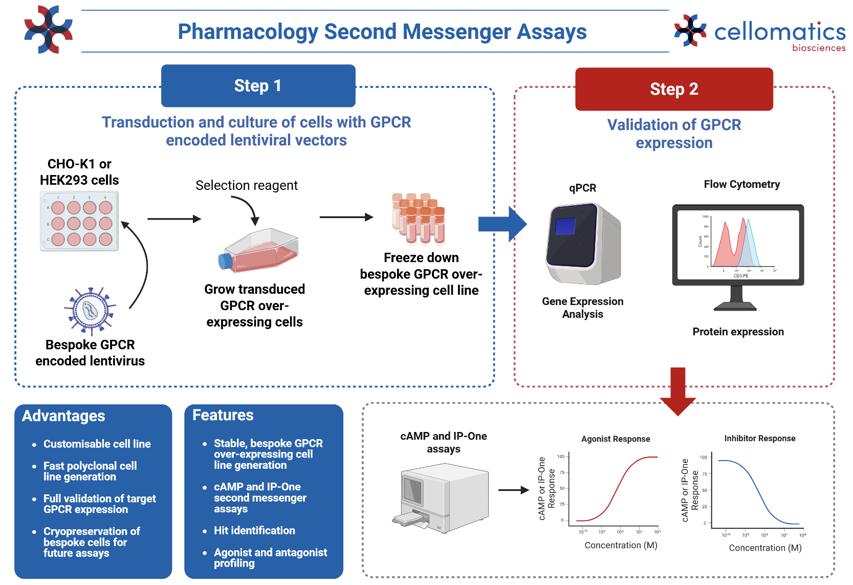
Verification of GPCR stable expression by qPCR and flow cytometry
To confirm the successful generation of GPCR-expressing stable cell lines, receptor expression can be verified at both the transcript and protein level. qPCR provides a quantitative assessment of GPCR mRNA expression, while flow cytometry can be used to confirm receptor expression at the cell surface and assess the proportion and relative expression level within the cell population. Together, these complementary approaches provide robust validation of GPCR stable expression prior to downstream functional pharmacology studies.
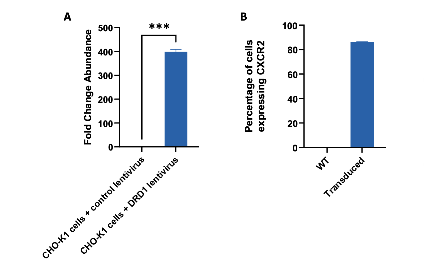
A) CHO-K1 cells transduced with DRD1-lentivirus express nearly 400-fold more DRD1 RNA than a negative control lentivirus. CHO-K1 cells were transduced with lentivirus with and without the DRD1 gene encoded using a multiplicity of infection of 20. Transduced CHO-K1 cells were maintained under puromycin selection, harvested for qPCR and analysed for expression of DRD1. Data were normalised to the negative control lentivirus and show mean n=1 ± SEM from three technical replicates. B) HEK293 cells transduced with CXCR2-lentivirus express CXCR2 in over 80% of cells, compared to 0% in wild-type. HEK293 cells were transduced with lentivirus with and without the CXCR2 gene encoded using a multiplicity of infection of 1. Transduced HEK293 cells were maintained under puromycin selection, harvested for flow cytometry and analysed for expression of CXCR2. Data show mean n=1 ± SEM from three technical replicates.
Agonist and antagonist assays of GPCR stably expressing cells for Gi, Gs and Gq signalling
Agonist and antagonist assays provide a functional assessment of GPCR activity and compound pharmacology across the major G-protein signalling pathways. Using stable GPCR-expressing cell lines, Gᵢ-, Gₛ- and Gq-coupled receptors can be evaluated through appropriate second-messenger readouts, including cAMP and IP-One assays. These assays enable determination of compound potency, efficacy and inhibitory activity, supporting the characterisation and profiling of GPCR agonists and antagonists.
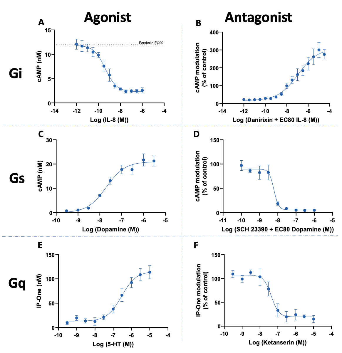
Agonist and antagonist profiling in GPCR stably expressing cells for Gi, Gs and Gq signalling. A and B) CXCR2 over-expressing HEK293 cells responded to a 30-minute treatment of IL-8 agonist (A) or Danirixin antagonist (B) in a concentration dependent manner. Pre-determined Forskolin EC90 concentration was used to stimulate adenylyl cyclase. Pre-determined IL-8 EC80 concentration was used to challenge the Danirixin antagonist. cAMP levels were quantified in a Gi assay and raw data was interpolated against a cAMP standard curve (n=3 ± SEM). C and D) DRD1 over-expressing CHO-K1 cells responded to a 30-minute treatment of dopamine agonist (C) or SCH23390 antagonist (D) in a concentration dependent manner. Pre-determined dopamine EC80 concentration was used to challenge the SCH23390 antagonist. cAMP levels were quantified in a Gs assay and raw data was interpolated against a cAMP standard curve (for (C) n=6 ± SEM; for (D) n=3 ± SEM). E) and F) 5-HT2A over-expressing CHO-K1 cells responded to a 90-minute treatment of 5-HT agonist (E) or 5-HT2A antagonist Ketanserin (F, blue) and CXCR2 antagonist, Danirixin (F, red). Pre-determined 5-HT EC80 concentration was used to challenge the antagonists. IP-One levels were quantified in a Gq assay and raw data was interpolated against an IP-One standard curve (for (E) n=4 ± SEM; for Ketanserin treatment, n=3 ± SEM; for Danirixin treatment, n=1 ± SEM).
Agonist and antagonist assays in GPCR stably expressing cells
Agonist and antagonist assays in GPCR stably expressing cells enable direct functional assessment of receptor activation and inhibition. Using validated cell lines with consistent receptor expression, concentration–response studies can be performed to determine compound potency, efficacy and inhibitory activity, including EC50 and IC50 values. These assays provide a robust and reproducible platform for GPCR compound screening, hit confirmation and pharmacological characterisation.
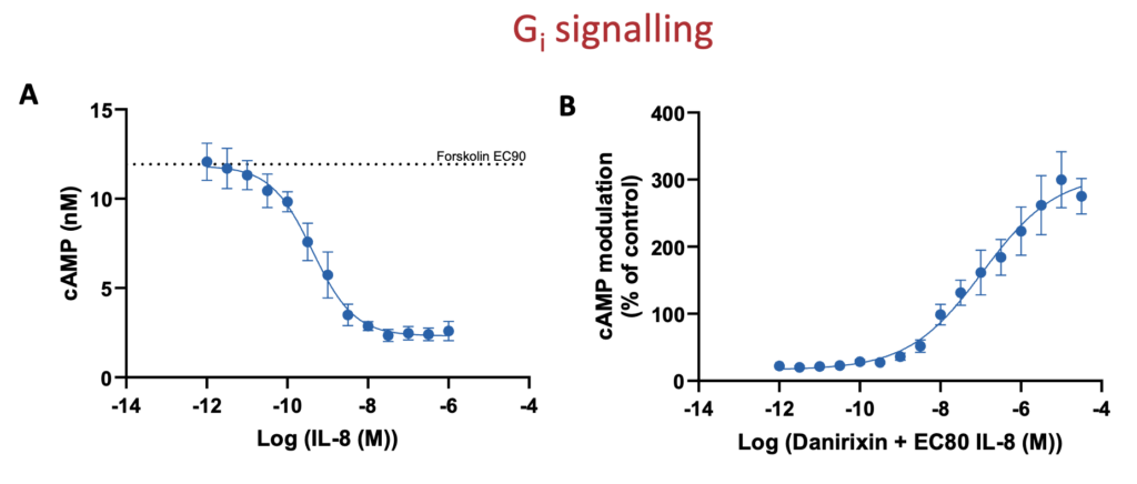
Agonist and antagonist assays in cells stably overexpressing CXCR2 GPCR for Gi signalling. A) CXCR2 over-expressing HEK293 cells responded to a 30-minute treatment of IL-8 agonist or B) Danirixin antagonist in a concentration dependent manner. Pre-determined Forskolin EC90 concentration was used to stimulate adenylyl cyclase. Pre-determined IL-8 EC80 concentration was used to challenge the Danirixin antagonist. cAMP levels were quantified in a Gi assay and raw data was interpolated against a cAMP standard curve (n=3 ± SEM).
Agonist and antagonist assays using GPCR stably expressing cells provide a reliable platform for functional pharmacology and compound profiling. By measuring receptor-mediated signalling in response to increasing concentrations of test compounds, these assays allow determination of potency, efficacy, EC50 and IC50 values, supporting hit identification, lead optimisation and comparative characterisation of GPCR modulators.
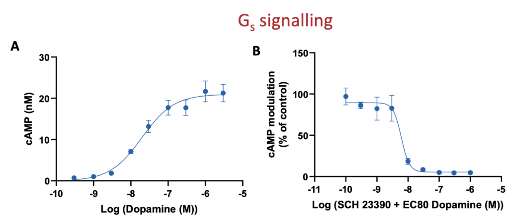
Agonist and antagonist assays in cells stably overexpressing DRD1 GPCR for Gs signalling. A) DRD1 over-expressing CHO-K1 cells responded to a 30-minute treatment of dopamine agonist or B) SCH23390 antagonist in a concentration dependent manner. Pre-determined dopamine EC80 concentration was used to challenge the SCH23390 antagonist. cAMP levels were quantified in a Gs assay and raw data was interpolated against a cAMP standard curve (for (A) n=6 ± SEM; for (B) n=3 ± SEM).
Agonist and antagonist assays in GPCR stably expressing cells provide a robust approach for assessing receptor-specific compound activity. These assays enable concentration–response profiling of test compounds to determine agonist potency and efficacy or antagonist inhibitory activity, supporting the calculation of key pharmacological parameters such as EC50 and IC50 values. This approach is well suited to GPCR compound screening, hit validation and comparative pharmacological characterisation.
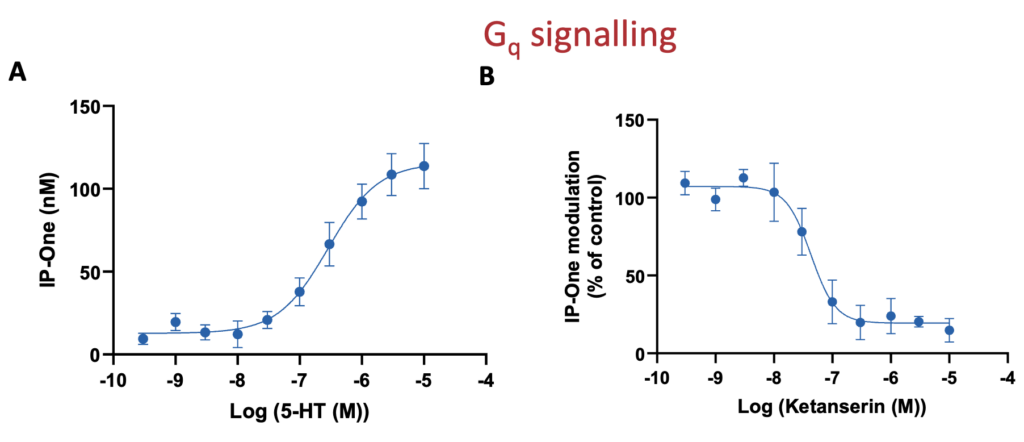
Agonist and antagonist assays in cells stably overexpressing 5-HT2A GPCR for Gq signalling. A) 5-HT2A over-expressing CHO-K1 cells responded to a 90-minute treatment of 5-HT agonist or B) Ketanserin antagonist in a concentration dependent manner. Pre-determined 5-HT EC80 concentration was used to challenge the ketanserin antagonist. IP-One levels were quantified in a Gq assay and raw data was interpolated against an IP-One standard curve (for (A) n=4 ± SEM; for (B) n=3 ± SEM).
β-arrestin assays in GPCR stably expressing cells
β-arrestin assays provide a complementary approach to classical G-protein signalling assays for characterising GPCR activity. Following receptor activation, β-arrestin recruitment can be measured to assess receptor desensitisation, internalisation and β-arrestin-mediated signalling. In GPCR stably expressing cells, these assays support agonist and antagonist profiling, potency determination, biased agonism studies and broader GPCR compound characterisation.
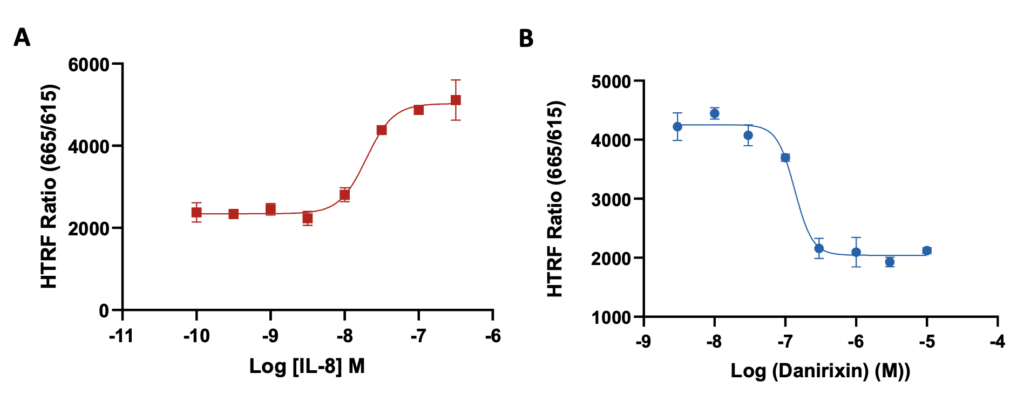
β–arrestin assays in cells stably overexpressing CXCR2 GPCR. CXCR2 over-expressing HEK293 cells were serum starved for 4 hours before a 30-minutes treatment of A) IL-8 agonist or B) Danirixin antagonist in a concentration dependent manner. Pre-determined IL-8 EC80 concentration was used to challenge the Danirixin antagonist. HTRF ratio was quantified using a β-arrestin assay (n=1 ± SEM).
Request a consultation with Cellomatics Biosciences today
Our experienced team of in vitro laboratory scientists will work with you to understand your project and provide a bespoke project plan with a professional, flexible service and a fast turnaround time.
To request a consultation where we can discuss your exact requirements, please contact Cellomatics Biosciences.