
Cell invasion is a finely regulated processes that is critical in many normal physiological functions including embryonic development, wound repair and immune surveillance (Lauffenburger and Horwitz, 1996; Pollard and Borisy, 2003; Ridley et al., 2003). However, these dynamic cell movements are also crucial in cancer progression and metastasis (Hamidi and Ivaska, 2018).
Here at Cellomatics, we support a wide range of projects with a focus on cell invasion (and migration) utilising well established invasion assays.
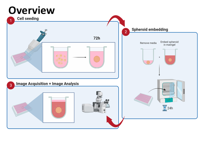
Principle
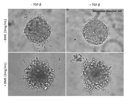
HT-1080 (human fibrosarcoma cell line) cells were grown as 3D spheroids for 72 hours before embedding in Basement Membrane Extract (BME) for 5-10 days in the presence or absence of TGFβ. Images were captured with a brightfield microscope – 10X
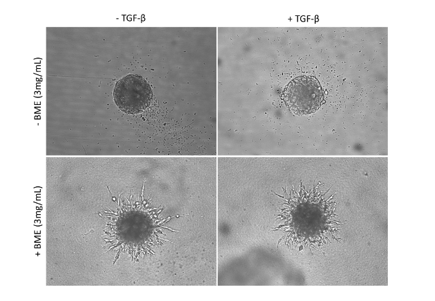
NIH/3T3 (mouse fibroblast cell line) cells were grown as 3D spheroids for 72 hours before embedding in Basement Membrane Extract (BME) for 5-10 days in the presence or absence of TGFβ. Images were captured with a brightfield microscope – 10X
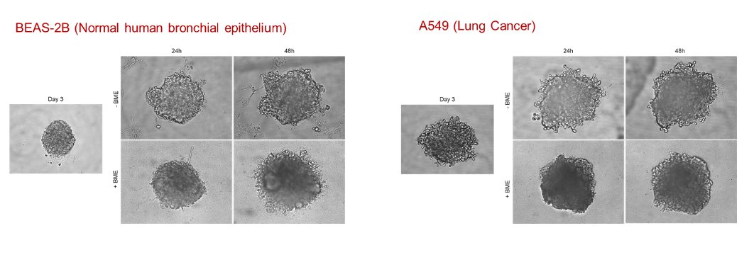
BEAS-2B (Normal human bronchial epithelium) and A549 (human lung epithelial cancer cell line) cells were grown as 3D spheroids for up to 72 hours with/without embedding in Basement Membrane Extract (BME) to study their invasive properties. Images were captured with a brightfield microscope – 10X
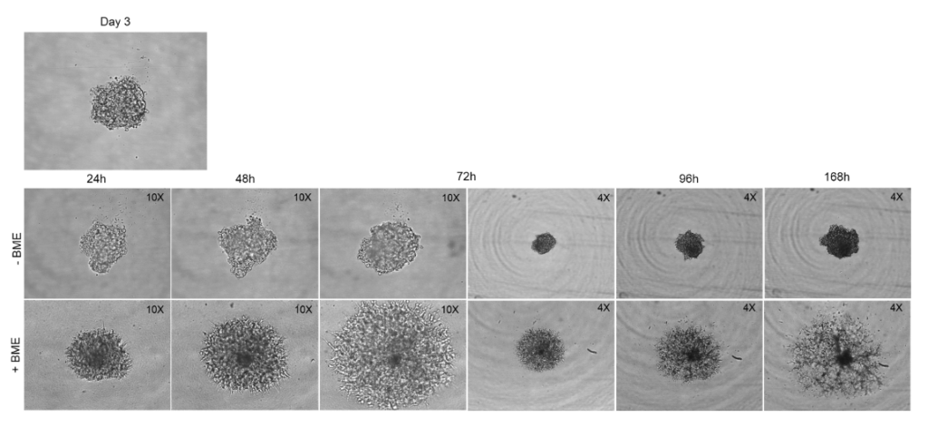
SH-5YSY (Neuroblastoma) cells were grown as 3D spheroids for up to 7 days with/without embedding in Basement Membrane Extract (BME) to study their invasive properties. Images were captured with a brightfield microscope – x10 and x4
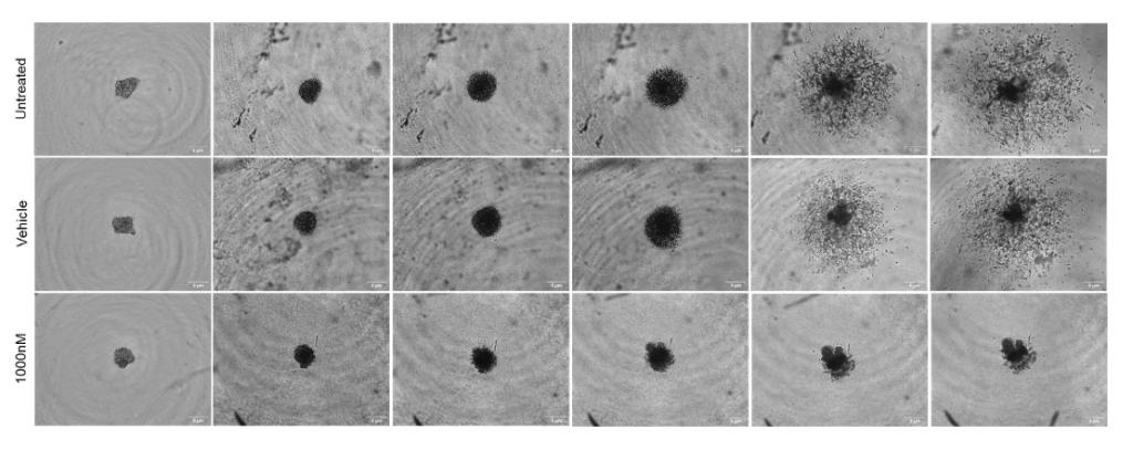
SH-5YSY (Neuroblastoma) cells were grown as 3D spheroids for up to 9 days by embedding in Basement Membrane Extract (BME) in the presence or absence of Cytochalasin D (potent inhibitor of actin polymerization). Images were captured with a brightfield microscope – 4X
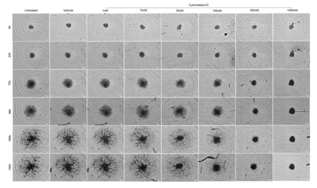
SH-5YSY (Neuroblastoma) cells were grown as 3D spheroids for up to 8 days by embedding in Basement Membrane Extract (BME) in the presence or absence of Cytochalasin D (potent inhibitor of actin polymerization) in a 6-concentration response curve. Images were captured with a brightfield microscope – 4X
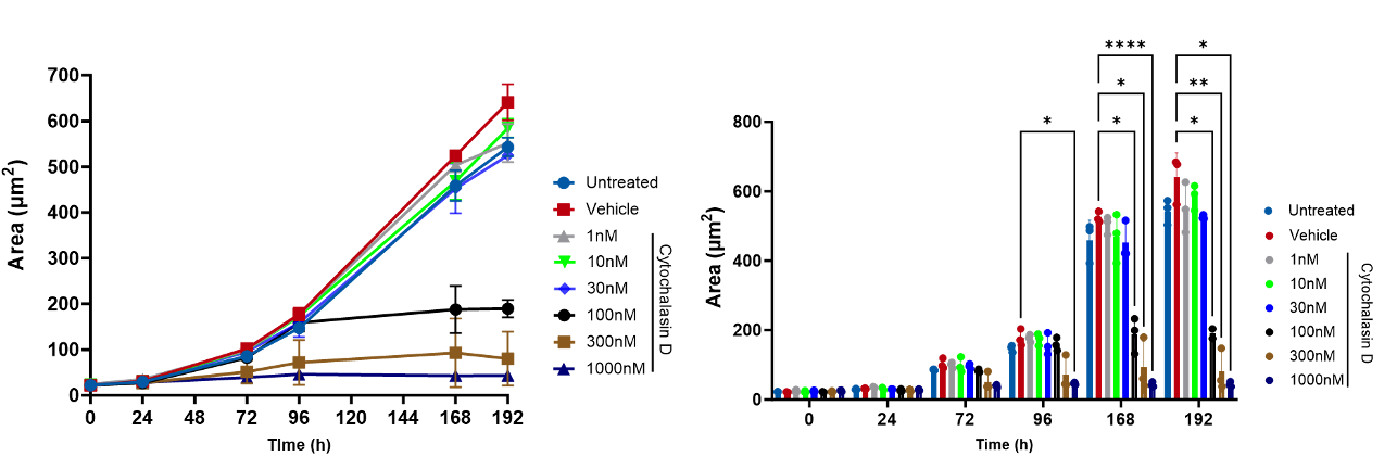
SH-5YSY (Neuroblastoma) cells were grown as 3D spheroids for up to 8 days by embedding in Basement Membrane Extract (BME) in the presence or absence of Cytochalasin D (potent inhibitor of actin polymerization) in a 6-concentration response curve. Images were captured with a brightfield microscope – 4X. Data analysed and plotted as total spheroid area (µm2)
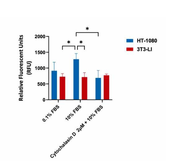
Invasion assay in Human Fibrosarcoma HT-1080 and mouse fibroblast 3T3-LI cells (negative control). Cells were seeded into Corning Transwell™ inserts and were coated with Cultrex® Basement Membrane Extract. Cells seeded in serum-free media invaded towards FBS for 48hrs in the presence or absence of Cytochalsain D. Invasion assay showed that treatment with 2µM Cytochalsain D resulted in a significant decrease in invasion with highly invasive HT-1080 cells, as indicated in the relative fluorescent values (fluorescence enhancement when bound to cellular nucleic acids; more cells more fluoresence).
Our experienced team of in vitro laboratory scientists will work with you to understand your project and provide a bespoke project plan with a professional, flexible service and a fast turnaround time.
To request a consultation where we can discuss your exact requirements, please contact Cellomatics Biosciences.
Cellomatics Biosciences Limited
10 Colwick Quays Business Park
Private Road No 2, Colwick Nottingham NG4 2JY, UK
+44 (115) 865 4101
info@cellomaticsbio.com

Cellomatics Biosciences Limited
10 Colwick Quays Business Park
Road No2, Colwick Nottingham NG4 2JY, UK
+44 (115) 865 4101
info@cellomaticsbio.com


| Cookie | Duration | Description |
|---|---|---|
| cookielawinfo-checkbox-analytics | 11 months | This cookie is set by GDPR Cookie Consent plugin. The cookie is used to store the user consent for the cookies in the category "Analytics". |
| cookielawinfo-checkbox-functional | 11 months | The cookie is set by GDPR cookie consent to record the user consent for the cookies in the category "Functional". |
| cookielawinfo-checkbox-necessary | 11 months | This cookie is set by GDPR Cookie Consent plugin. The cookies is used to store the user consent for the cookies in the category "Necessary". |
| cookielawinfo-checkbox-others | 11 months | This cookie is set by GDPR Cookie Consent plugin. The cookie is used to store the user consent for the cookies in the category "Other. |
| cookielawinfo-checkbox-performance | 11 months | This cookie is set by GDPR Cookie Consent plugin. The cookie is used to store the user consent for the cookies in the category "Performance". |
| viewed_cookie_policy | 11 months | The cookie is set by the GDPR Cookie Consent plugin and is used to store whether or not user has consented to the use of cookies. It does not store any personal data. |