Inflammation Assays
Non-Alcoholic Steatohepatitis (NASH)
NASH is a condition that begins with fat build up around the liver, known as steatosis. The fat build up gets progressively worse and leads to inflammation and fibrosis of the liver. In turn, the disease progression eventually results in irreversible damage to the hepatic tissues and can lead to cirrhosis or hepatocellular carcinomas.
Causes and Risk Factors:
- Obesity
- Diabetes
- High Cholesterol
- Poor Diet
At present, no treatments exist for NASH and lifestyle and nutritional changes are recommended. The absence of available therapies is likely due to the lack of reliable in vitro models for this disease. However, significant progress has been made within this disease area, and Cellomatics is at the forefront of this research with a variety of NASH models available.
In Vitro Models
- Steatosis
- Fibrosis
- Metabolic Stress
- Inflammation
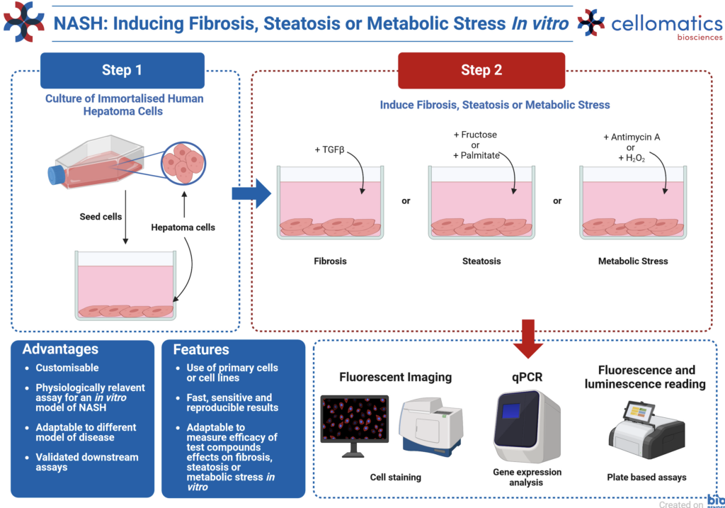
Fibrosis, Steatosis and Metabolic Stress
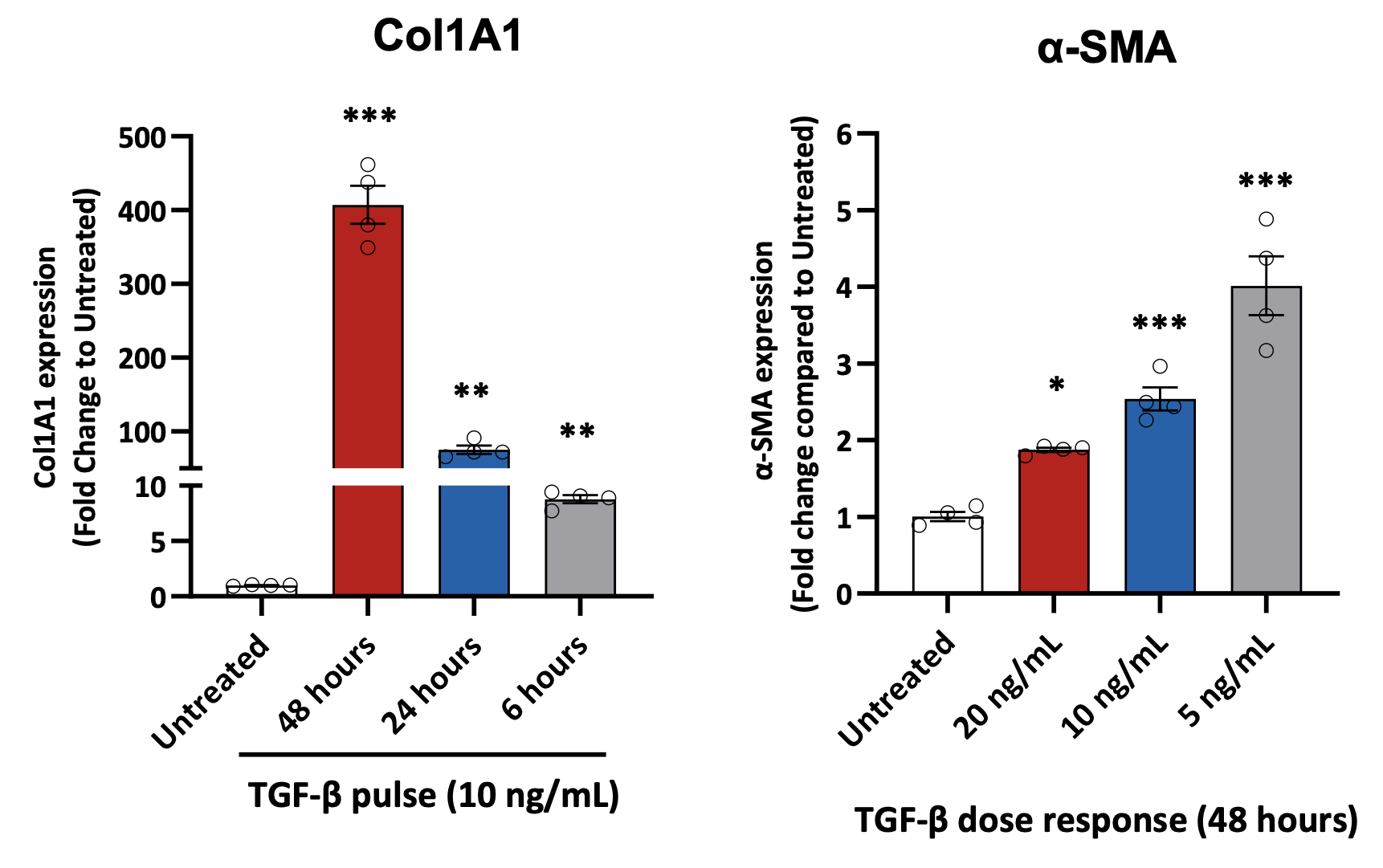
Fibrosis – Gene Expression Analysis
Fibrotic markers in HepG2 cells stimulated with TGF-β. Gene expression analysis of collagen type I alpha 1 (COL1A1) and α-smooth-muscle actin (α-SMA) in HepG2 cells stimulated with TGF-β. Data are expressed as means ± SEM; p˂0.05 considered statistically significant.
Steatosis – Lipid Quantification
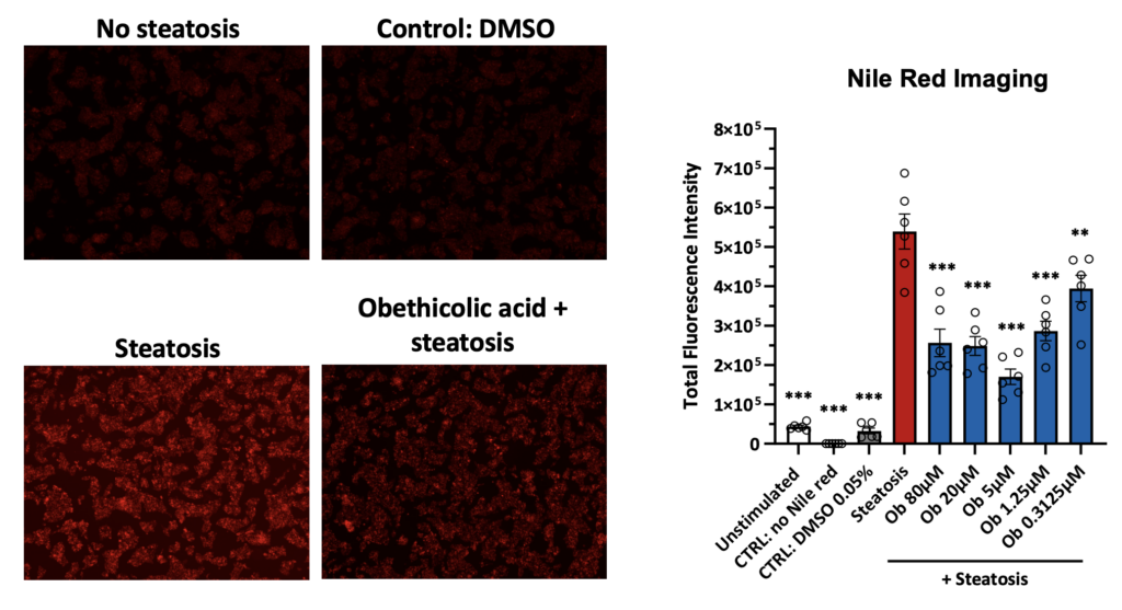
Induction of steatosis in HepG2 cells and Nile red staining quantification by imaging. Representative images of Nile red staining in HepG2 cells following induction of steatosis. HepG2 cells were incubated for 24 hours in relevant cell culture medium containing 100 mM of fructose and 100 µM of BSA-palmitate to induce steatosis. Different concentrations of Obethicolic acid (Ob), were used as a reference/tool compound. Data are expressed as means ± SEM; p˂0.05 considered statistically significant.
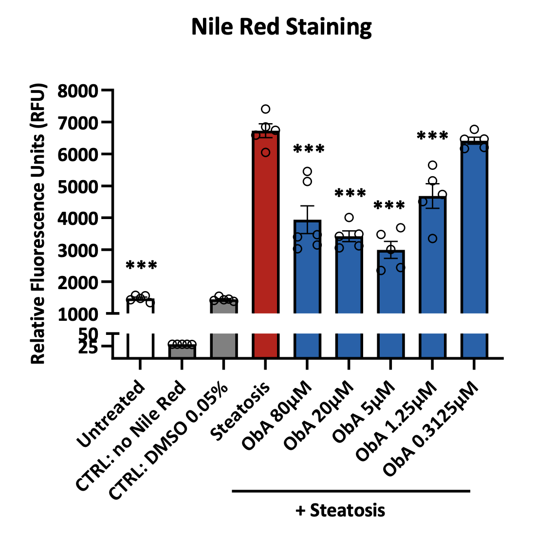
Induction of steatosis in HepG2 cells and quantification of Nile red staining by plate reader. HepG2 cells were plated in a 96 well plate and pre‑incubated with obethicolic acid (ObA) at the indicated concentrations before proceeding with the induction of steatosis for 24 hours. The potency of the obethicolic acid in reducing intracellular lipids was assessed measuring the fluorescence of Nile red staining on a plate reader. Data are expressed as means ± SEM; p˂0.05 considered statistically significant
Metabolic Stress – ROS Quantification
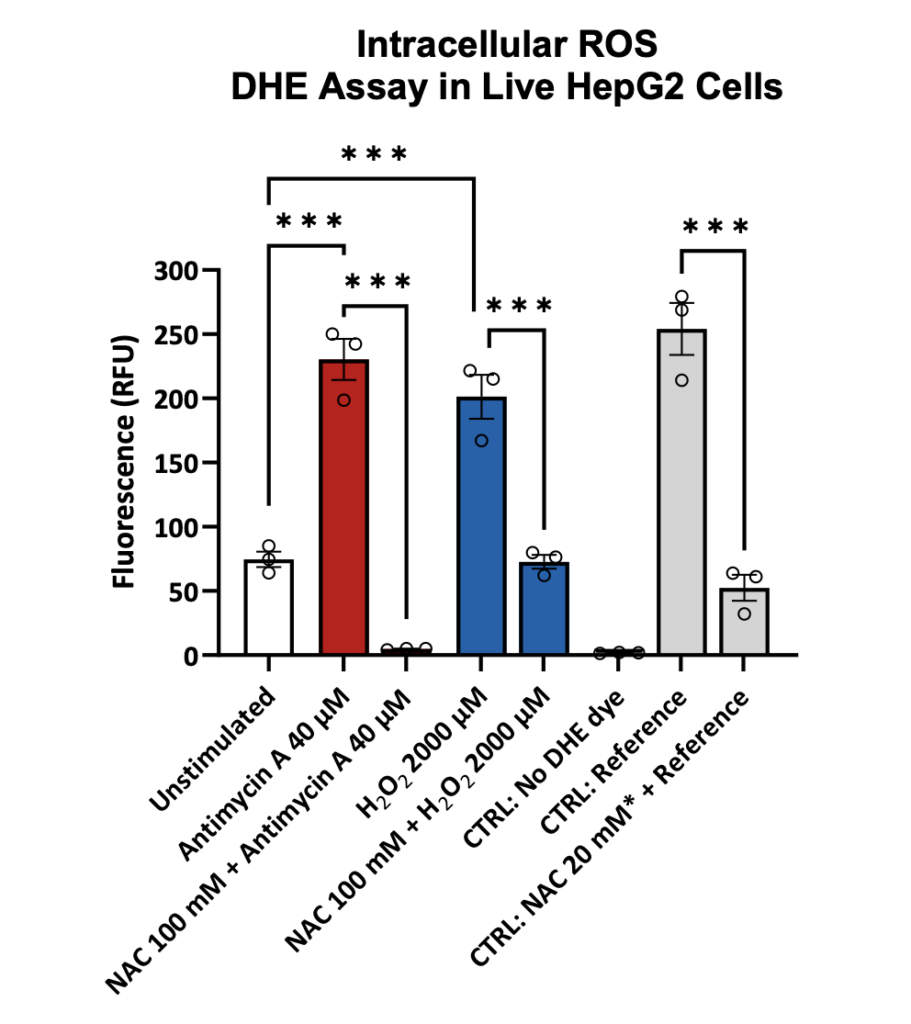
Levels of intracellular reactive oxygen species in HepG2 cells. Quantification of ROS in HepG2 cells following oxidative stress induced by validated ROS inducers (antimycin A and oxygen peroxide – H2O2). Data are expressed as means ± SEM. One-way ANOVA, p˂0.05 considered statistically significant (Reference = antimycin A standard, NAC = N‑acetylcysteine).
Inflammation
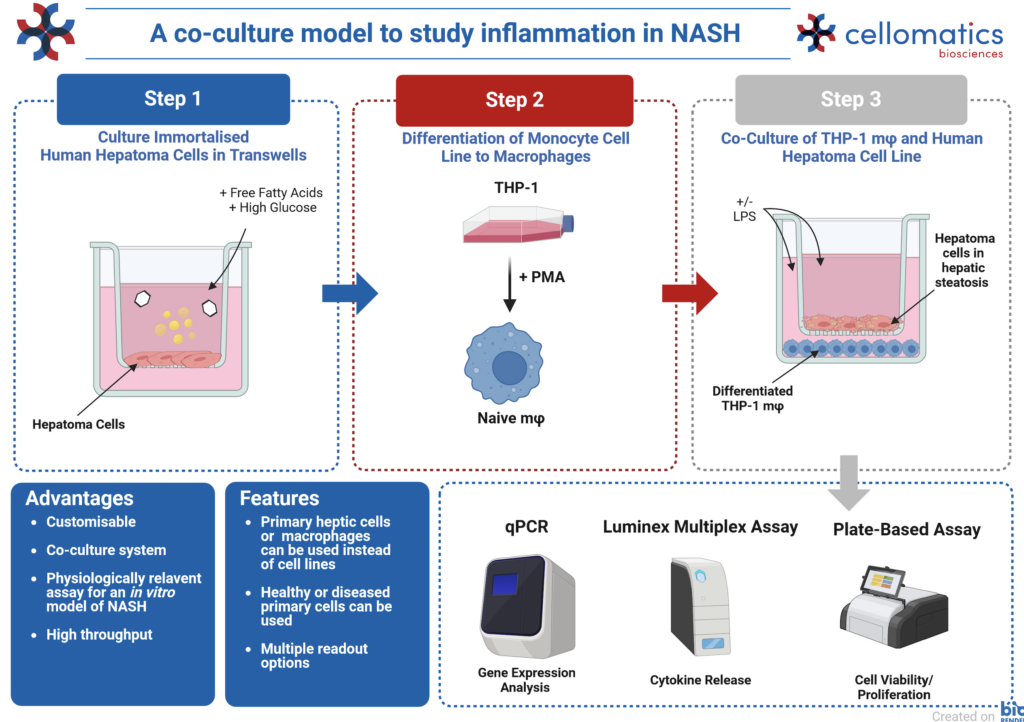
Cytokine Release in Inflammation
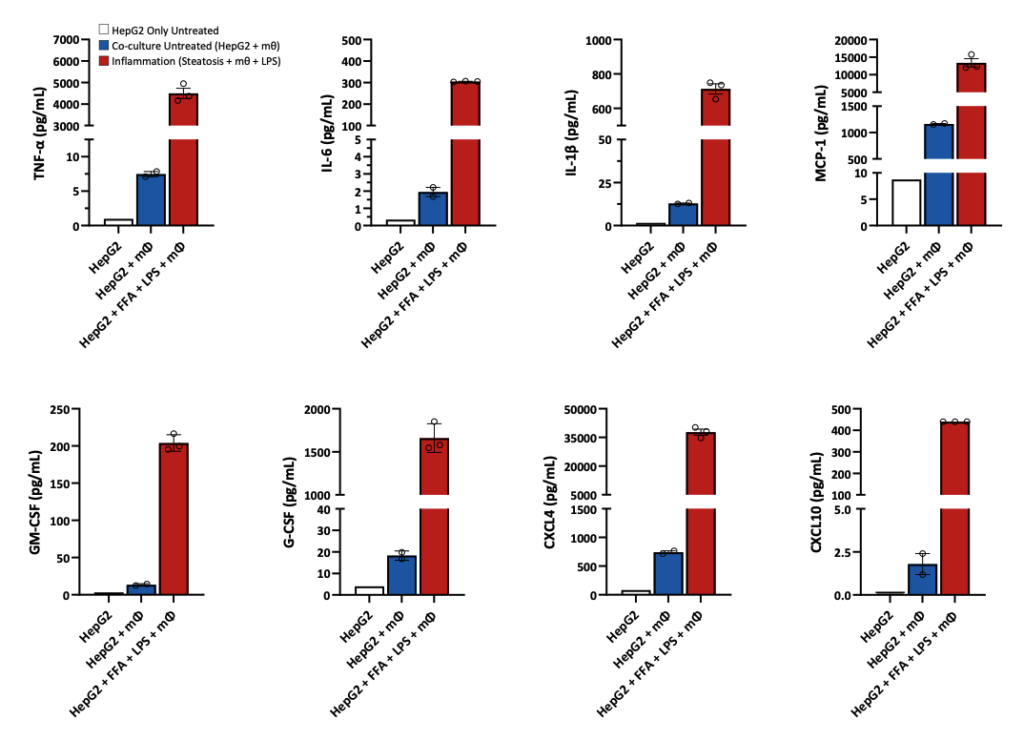
Quantification cytokines released in a pro‑inflammatory model of HepG2 and macrophages. HepG2 cells were seeded in the apical transwell of 24 well plate and steatosis induced with Oleic acid and BSA‑palmitate (FFA) according to our optimised protocol (Kanmani P et al. Frontiers in Immunology 2018). Co‑culture with polarised THP‑1 cells was initiated and inflammation induced by exposing the cells to LPS (1 µg/mL) for 24 hours. Supernatants were harvested and quantification of cytokines assessed by Luminex (xMAP). Data are expressed as means ± SEM.
FGF Quantification in Steatosis Induced Inflammation
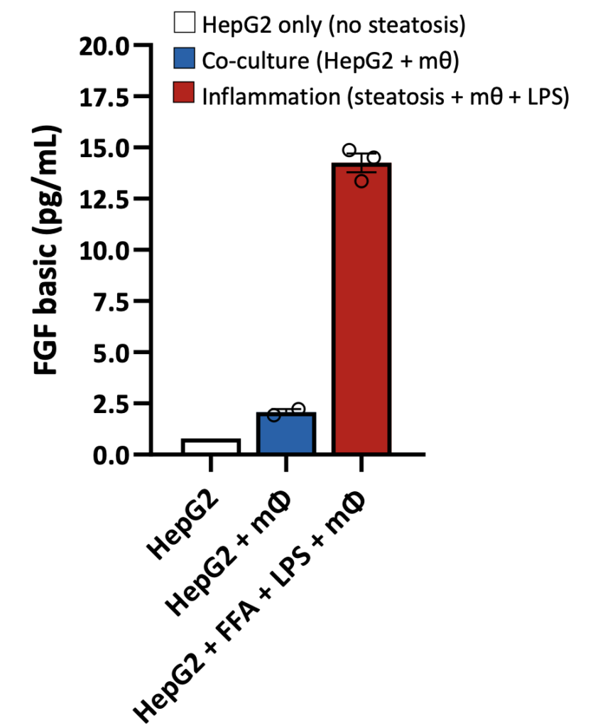
Quantification of FGF in the co‑culture model of HepG2 and macrophages. HepG2 cells were seeded in the apical transwell of 24 well plate and steatosis induced with Oleic acid and BSA‑palmitate (FFA) according to our optimised protocol (Kanmani P et al. Frontiers in Immunology 2018). Co‑culture with polarised THP‑1 cells was initiated and inflammation induced by exposing the cells to LPS (1 µg/mL) for 24 hours. Supernatants were harvested and quantification of cytokines assessed by Luminex (xMAP). Data are expressed as means ± SEM.
Request a consultation with Cellomatics Biosciences today
Our experienced team of in vitro laboratory scientists will work with you to understand your project and provide a bespoke project plan with a professional, flexible service and a fast turnaround time.
To request a consultation where we can discuss your exact requirements, please contact Cellomatics Biosciences.


